Radiation CT radiographing device, radiation CT radiographing system, and radiation CT radiographing method using the same

a radiographing device and radiographing system technology, applied in the direction of tomography, patient positioning for diagnostics, instruments, etc., can solve the problems of object dizziness, centrifugal force applied to the object also increases, and the photographing fails, etc., to achieve high processing speed, low cost, and high sensitivity

Inactive Publication Date: 2007-12-18
CANON KK
View PDF30 Cites 111 Cited by
  • Summary
  • Abstract
  • Description
  • Claims
  • Application Information

AI Technical Summary

Benefits of technology

[0034]The invention is made in consideration of the foregoing conventional problems and it is an object of the invention to provide an object rotating type cone beam radiation CT radiographing device and a radiation CT radiographing system in which a high processing speed and high sensitivity are obtained with low costs and to provide a radiation CT radiographing method using such a device and a system. Thus, a radiation CT device of high performance for a vehicle-mounted use or a group medical examination can be provided at low costs.
[0046]In the invention, since there is no need to wait until the rotation becomes stable and the wasteful rotation time can be shortened, it is comfortable for the patient. The dedicated reference element is unnecessary. Since the panel can be used as it is, the system is simplified and the costs are reduced. Further, there is no need to prepare the reference element on the X-ray source side. The X-ray source for the general photographing can be used in common. Since it is sufficient to arrange the rotating device and the X-ray image sensor in the general photographing room, the expenses of the facilities can be reduced. Since it is sufficient to use the rotating device with the general mechanism, the costs can be easily reduced. Even if there is a slight rotational variation, it is possible to cope with it. The mechanism for accurately controlling the rotation is unnecessary. Since the system construction is simple, the maintenance is easy to perform and it is convenient to mount the system onto the general examination vehicle.
[0047]According to the invention, the object rotating type cone beam radiation radiographing device having high processing speed, high sensitivity, and high performance with low costs can be realized by the X-ray image sensor panel of the large panel size using a plurality of CMOS type image pick-up elements and by the novel driving method. Thus, the radiation CT radiographing device of high performance for a vehicle-mounted use and group medical examination can be provided at low costs.

Problems solved by technology

There is a problem that if the object is rotated at a speed higher than such a rotating time, the object enters what is called a dizzy state where he feels dizzy.
There is also a problem that when the object is rotated at a high speed, a centrifugal force which is applied to the object also increases together with an increase in rotating speed, so that the object is moved during the measurement.
There is also a problem that since the breath pausing time of about 5 seconds per rotation is relatively long, an examinee feels anxiety during the examination, a body movement artifact occurs due to an unexpected operation, body vibration, or the like, or the object is out of a field of vision of the detector, so that the photographing fails.
There is also a problem that if the object is moved during the measurement, “blur” or “deflection” occurs in a 3-dimensional X-ray distribution image formed by the reconstruction and picture quality of a 3-dimensional X-ray image deteriorates.
There is, consequently, a problem that more than one rotation is necessary and a burden on the patient becomes heavy.
There is also a problem that if the photographing is started in the unstable state, rotational angles of respective projections differ, precision of the reconstruction deteriorates, a variation in a signal from a reference element increases, precision of the correction deteriorates, and the image deteriorates.
There is also a problem that, generally, in the X-ray CT radiographing device, even if the X-ray is radiated while a lamp voltage and a lamp current are set to the same conditions, upon photographing, X-ray intensity actually changes for every scanning, in detail, and in every viewing.
This problem is caused by focal position changes or the like due to expansion / contraction of a shaft of an anode by a temperature change of an X-ray valve, movement of the shaft between bearings due to the motion of the valve, precession motion when the anode is rotating, and the like.
However, there is also a similar problem in the object rotating type cone beam X-ray CT radiographing device.
There is also a problem that a complicated mechanism and control apparatus are necessary to rotate the rotating plate on which various objects are mounted while it is precisely controlled, so that the manufacturing costs rise.
There is also such a problem that in the case where the rotating plate on which the object is mounted is photographed while purposely changing the rotating speed in dependence on the photographing region of the object, an accurate image of each region cannot be obtained corresponding to each rotating speed set for every photographing region of the object.
However, there is such a problem that it is very difficult to drive the flat panel detector using a thin film transistor of a-Si or a-Se with an area of the 17-inch size (43 cm×43 cm) or more at such a speed because semiconductor characteristics of a-Si or a-Se are inadequate.
There is such a problem that in the cone beam X-ray CT radiographing device in which an X-ray dosage per projection is fairly smaller than that of the still image, since noises are large and the sensitivity is low, the sensitivity in the conventional flat panel detector is insufficient.
That is, there are the following problems: an essential problem in which a distortion of the image occurs in the peripheral portion; a problem in which even in the current maximum 17-inch size, since the display screen is circular, an wide enough area almost equal to that of the flat panel detector cannot be obtained, the reconstructing area decreases remarkably in the cone beam X-ray CT; and a problem in which, since the magnetism is used for controlling an electron beam, if the device is rotated, it is complicatedly influenced by the terrestrial magnetism.
However, the following problems of the X-ray source exist.
Since such a phenomenon is caused due to a temperature change, aging change, abrasion of the electrode, or the like, it is impossible to avoid the situation wherein the focal position differs by a difference of the time position upon collection of X-ray absorption data, that is, by a difference of the X-ray exposing time point.
A fluctuation of sensitivity distribution of the detector due to the movement of the focal point becomes a calibration error resulting in an artifact on the reconstructed image.
However, in the case of rotating the object at a high speed, the object enters what is called a dizzy state where he feels dizzy.
Naturally, the X-ray has to be radiated from the X-ray source for such a period of time and the focus movement mentioned above becomes a problem.
Moreover, since a countermeasure against the continuous photographing of a long period of time is not taken in the X-ray source for the general photographing, such a problem is more serious than that in the case of the X-ray source only for the X-ray CT radiographing.
There is also such a problem that when the existing X-ray source is used, there are various histories and the change in age, abrasion of the electrode, and the like, which also differ with every device, so that it has to cope with them for every X-ray source.
Further, since the X-ray source for the general photographing is not equipped with the foregoing reference detector, the reference detector has to be separately prepared and installed in the case of constructing the system.

Method used


Image

  • Radiation CT radiographing device, radiation CT radiographing system, and radiation CT radiographing method using the same
  • Radiation CT radiographing device, radiation CT radiographing system, and radiation CT radiographing method using the same
  • Radiation CT radiographing device, radiation CT radiographing system, and radiation CT radiographing method using the same

Examples


first embodiment

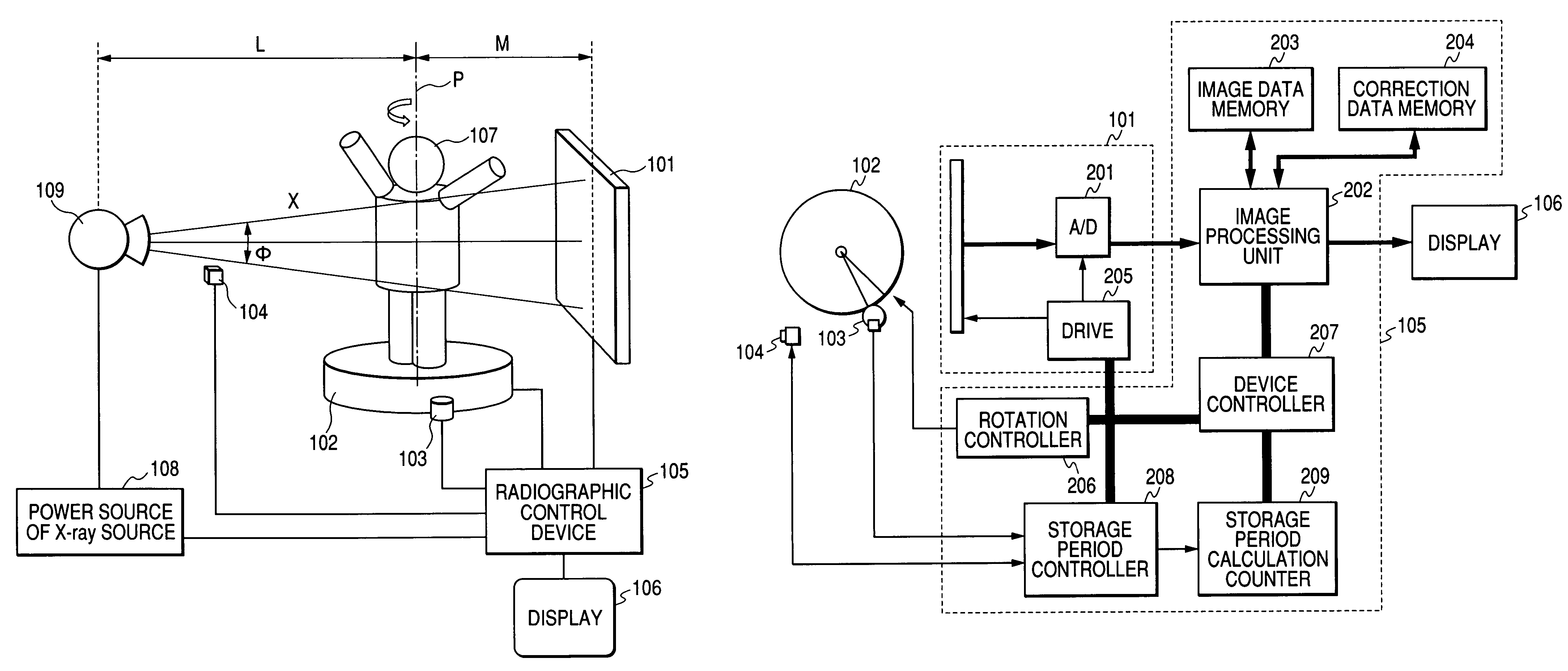
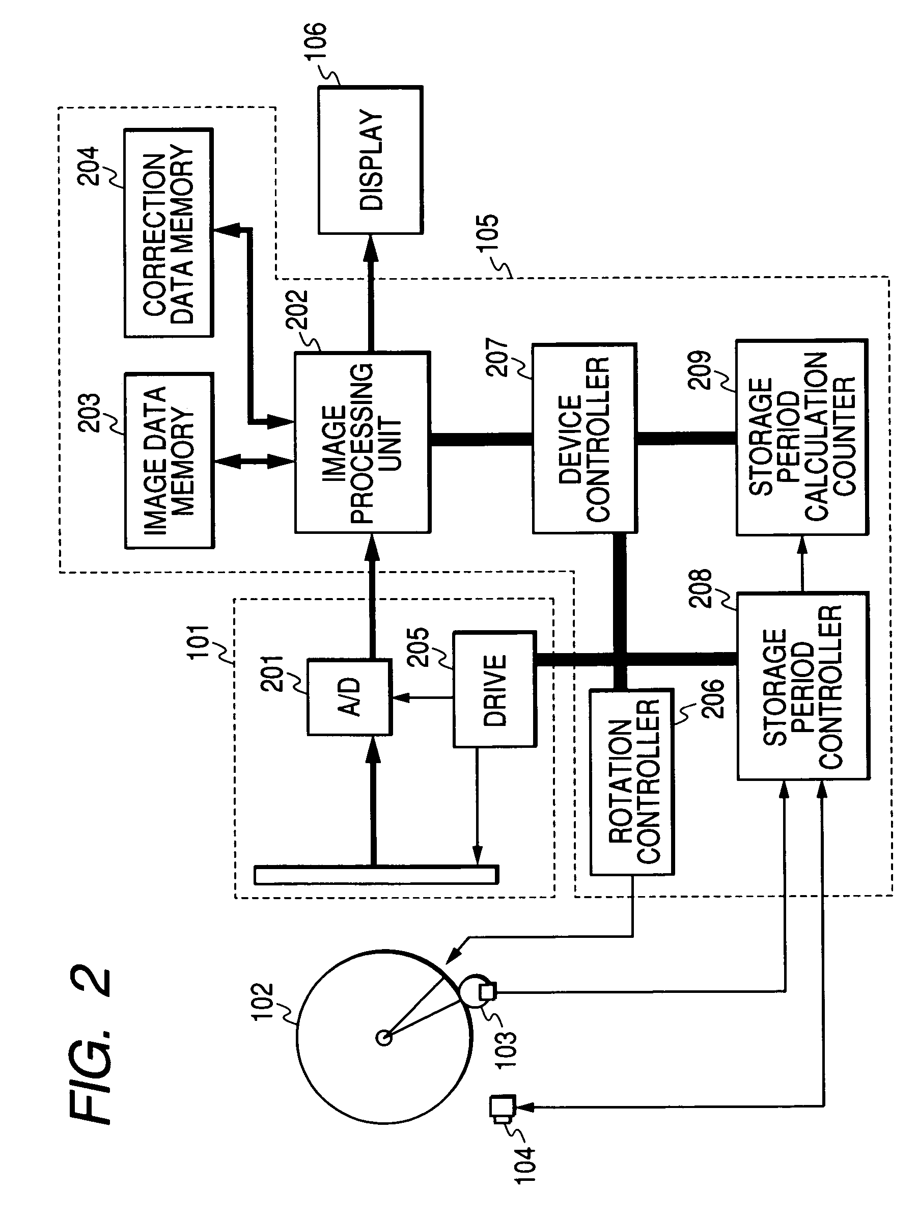
[0070]FIG. 1 is a diagram for explaining a schematic construction of the first embodiment of an object rotating type cone beam radiation CT radiographing device according to the invention. In the diagram, reference numeral 109 denotes an X-ray generator (X-ray source); 108 a power source for an X-ray source; X a radiated continuous X-ray; 107 an object; 104 a signal storage type reference element; 102 a rotating device (rotating plate); P a rotary axis of the rotating device; 103 a rotary encoder; 101 an X-ray image sensor panel; 105 a radiographic control device; and 106 a display. Although the X-ray is used as a radiation in the embodiment, another radiation such as α-ray, β-ray, γ-ray, or the like can be used. This is true of all embodiments.

[0071]The X-ray generator 109 is used for general photographing and is used in a continuous X-ray mode. The radiographic control device 105 sets a lamp voltage, a lamp current, and radiation time and controls only the start and stop of the ra...

second embodiment

[0132]According to the second embodiment, an ionization dosimeter is used as a signal storage type reference element. Generally, the ionization dosimeter (area dosimeter) is used as a measuring instrument which is attached to an X-ray radioscopic device and measures radiation exposure of the patient at the time of the radioscopic operation. The area dosimeter is attached to a movable diaphragm portion of the X-ray generator. Component elements other than the area dosimeter are similar to those in the first embodiment.

[0133]In the second embodiment, the area dosimeter is set so as to cover the front surface of the movable diaphragm of the X-ray generator. A value which is measured by the area dosimeter is a total dosage of the X-ray which is generated from a range limited by the movable diaphragm in the X-ray which is generated from the X-ray generator. In the first embodiment, the signal storage type reference element is set to a position where it does not become a shadow on the obj...

third embodiment

[0135]According to the third embodiment, a part of the X-ray image sensor panel is used as a reference element. The operation of the X-ray image sensor panel 101 is substantially the same as that of the first embodiment. As shown in FIG. 4, an X-ray radioscopic image includes portions without a projection image of the object, that is, blank portions. A region 401 surrounded by a broken line among the blank portions is used as a reference signal detecting portion. This region is determined by radiating the very weak X-ray prior to the main scanning and detecting the blank portions. After the reference signal detecting portion is determined, a collimator for removing scattered lines from the object is set to the relevant location of the X-ray image sensor panel. A grid can be also used in common. In the embodiment, since another reference detecting element is unnecessary, the image of the object and the reference signal can be simultaneously obtained as one image data. Thus, the syste...


PUM

PropertyMeasurementUnit
sizeaaaaaaaaaa
sizeaaaaaaaaaa
timeaaaaaaaaaa

Abstract

An object rotating type cone beam radiation CT radiographing device in which a high speed and high sensitivity are obtained with low costs is provided. Thus, a radiation CT device of high performance for vehicle-mounted use and group medical examination can be provided at low costs. In the object rotating type cone beam radiation CT radiographing device, a rotational angle of an object is detected, a signal storage period of a batch-exposure type radiation image sensor panel is controlled on the basis of the rotational angle signal, and a signal storage period of a signal storage type reference signal generator is controlled. There is provided a radiation CT radiographing device in which the object can be photographed at arbitrary resolution during the photographing, a fluctuation of a radiation source during the photographing can be detected without using a special detector and corrected in accordance with its detection amount.

Description

BACKGROUND OF THE INVENTION[0001]1. Field of the Invention[0002]The invention relates to a radiation CT (Computed Tomography) radiographing device for tomography photographing an object, a radiation CT radiographing system, and a radiation CT radiographing method using such a device and a system. More particularly, the invention relates to a cone beam radiation CT radiographing device using a 2-dimensional radiation image sensor panel. Further, the invention relates to a cone beam radiation CT radiographing device of an object rotating type by which an object such as an organism or the like in the state of standing on a floor or sitting on a chair is rotated and photographed between a radiation source and a radiation image sensor facing each other.[0003]2. Related Background Art[0004]In recent years, digitization has been progressed in various medical fields. Also in the X-ray diagnosis field, to digitize an image, there has been developed a 2-dimensional X-ray image pick-up device ...

Claims


Application Information

Patent Timeline
Patent Type & Authority Patents(United States)
IPC IPC(8): G01N23/00H05G1/60A61B6/03A61B6/04
CPCA61B6/032A61B6/04A61B6/547A61B6/4021A61B6/4291A61B6/0457A61B6/5258A61B6/0487
Inventor TASHIRO, KAZUAKIKAIFU, NORIYUKINAKANO, MIKIO
Owner CANON KK
Why Eureka
Try Eureka