Human immunosuppressive protein

a protein and immunosuppression technology, applied in the field of immunosuppression proteins, can solve the problems of limited viability, inability to meet the needs of patients, etc., and achieve the effect of suppressing the proliferation of responders, suppressing the production of t-cell proliferation and il-2, and reducing the viability

Inactive Publication Date: 2008-06-17
SOUTH FLORIDA UNIVESITY OF
View PDF18 Cites 12 Cited by
  • Summary
  • Abstract
  • Description
  • Claims
  • Application Information

AI Technical Summary

Benefits of technology

[0014]In another embodiment, there is a method of treating inflammation, the method comprising administering an effective amount of an immunosuppressant protein (HISP).
[0015]In yet another embodiment, there is an isolated immunosuppressant protein (HISP), the protein comprising an anionic protein and having a molecular weight of 40-100 kDa; having an isoelectric point of about 4.8; being obtained from hNT cell supernatant; not being obtained from NCCIT embryonal carcinoma cells, T98G glioblastoma cells or THP-1 monocytic leukemia cells; losing activity when treated with heat, pH 2, pH 11, or mixed with trypsin or carboxypeptidase; losing no acti

Problems solved by technology

Embryonic neurons as grafts are limited by their heterogeneity, expense, scarcity, diminishing viability over time, and refractoriness to standard transfection techniques (Cook et

Method used


Image

  • Human immunosuppressive protein
  • Human immunosuppressive protein
  • Human immunosuppressive protein

Examples


example 1

Production of Ntera2 / D1

[0071]Ntera2 / D1 cells (Layton Bioscience, Inc., Sunnyvale, Calif.) were maintained in Dulbecco's minimal essential medium with nutrient mixture F-12 (DMEM / F-12) with 2 mM L-glutamine, 100 U / ml penicillin, 100 μg / ml streptomycin, and 10% (v / v) heat-inactivated fetal bovine serum (FBS) (Life Technologies, Gaithersburg, Md.), and incubated in a 37° C., humidified, 5% CO2 environment.

example 2

Differentiation to hNT Neurons

[0072]Differentiation of Ntera2 / D1 is described in detail by Andrews, 1984 and Pleasure et al., 1992, which are hereby incorporated by reference. Briefly, Ntera2 / D1 (2×10⁶) were treated with 10 μM all-trans retinoic acid (Sigma, St. Louis, Mo.) for 5-6 weeks. Mitotic non-neuronal cells were inhibited with 1 μM cytosine β-D-arabinofuranoside, 10 μM 5-fluoro-2′-deoxyuridine, and 10 μM 1-β-D-ribofuranosyluracil (Sigma) for 1 week. Some retinoic acid-treated Ntera2 / D1 cultures were exposed to 25 Gy γ-irradiation (137Cs) to inhibit non-neuronal cells and were not treated with mitotic inhibitors, to ensure that trace amounts of mitotic inhibitors were not contributing to the immunosuppressive properties of hNT supernatant. Differentiated hNT neurons overlying the mixed cell culture were treated with 0.025% trypsin and 0.01% EDTA, dislodged, and replated at a density of 2-3×10⁶ cells / ml in serum-free Opti-Mem medium (Gibco-BRL), or DMEM / F-12 10% FBS medium, ea...

example 3

MHC Genotype & Phenotype

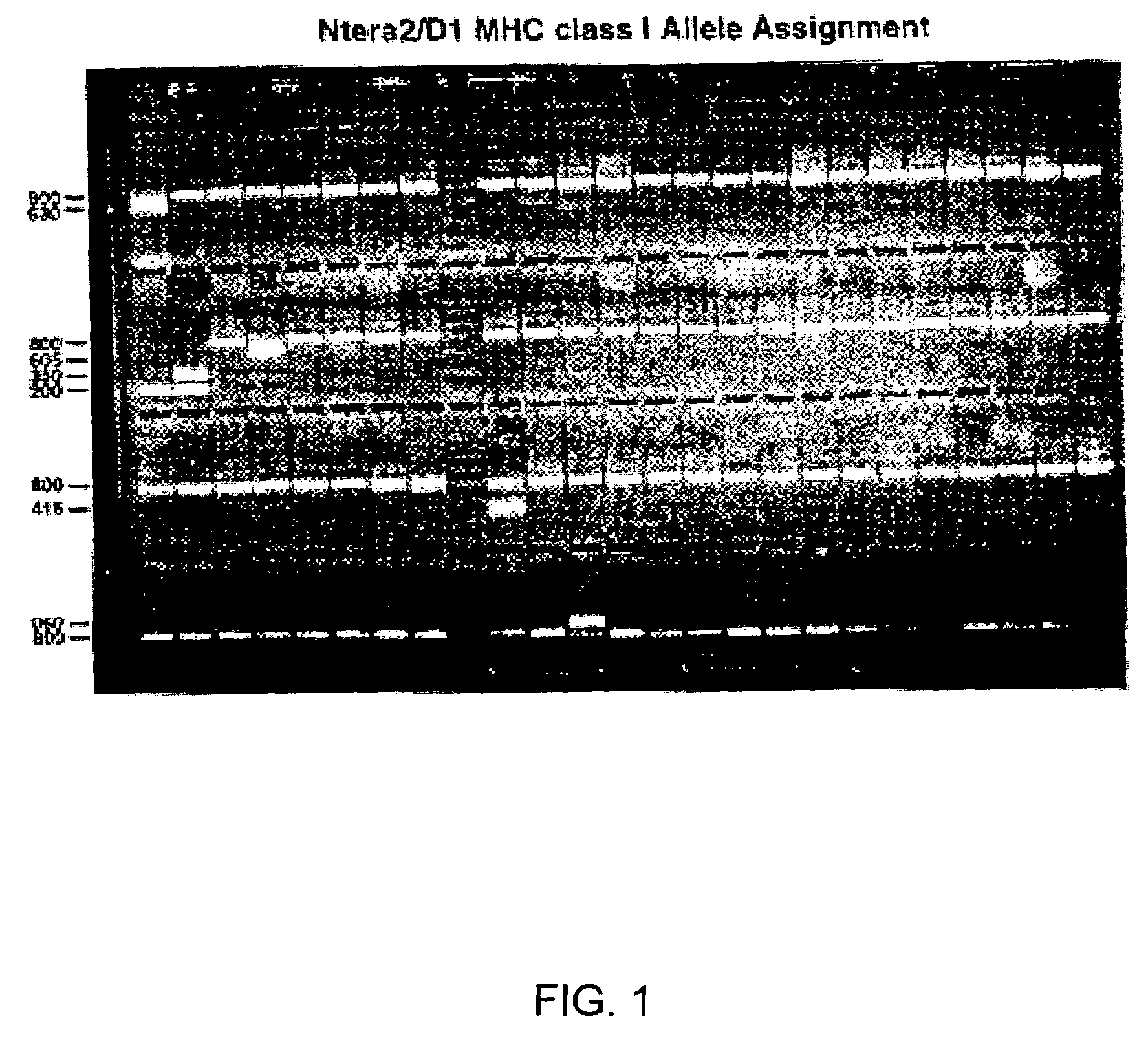
[0076]Ntera2 / D1 genomic DNA (75-125 ng / μl) was used as template for MHC class I and II analysis by sequence-specific primer polymerase chain reaction (Bunce et al., 1995) in MHC diagnostic plates (Pel-Freez Clinical Systems, Deerbrook Trail, Wis.), and the amplified products analyzed by ethidium bromide stained electrophoresis.

[0077]hNT MHC surface expression was detected using a complement-dependent cytotoxic technique in Terasaki tissue typing trays (One Lambda, Inc., Canoga Park, Calif.). Briefly, 2×10⁶ / ml hNT neurons were reacted with MHC antigen-specific monoclonal antibody (mAb) or known antisera, mixed with rabbit complement, ethidium bromide and acridine orange, and the reaction stopped by hemoglobin-EDTA. Cell viability was scored, and the MHC phenotype determined. At least 2 mAb or 3 overlapping antisera were used to define each MHC antigen.

[0078]The hNT neurons express MHC class I proteins. The amplified products of polymerase chain reactions using...


PUM

PropertyMeasurementUnit
Atomic weightaaaaaaaaaa
Fractionaaaaaaaaaa

Abstract

A method for purifying an immunosuppressant protein (HISP) has the steps of obtaining supernatant from hNT cells; exposing the supernatant to preparative polyacrylamide gel electrophoresis to produce 20 isoelectric fractions, including active isoelectric fraction #10; placing the active isoelectric fraction on a Blue Sepharose column to bind albumin; and collecting the free fraction containing the concentrated, isolated HISP. Also disclosed is a method of treating inflammation, using an effective amount of an HISP. The HISP is anionic, has a molecular weight of 40-100 kDa, an isoelectric point of about 4.8 and is obtained from the supernatant of hNT cells, but not from NCCIT embryonal carcinoma cells, T98G glioblastoma cells or THP-1 monocytic leukemia cells. HISP can maintain T cells in a quiescent G0/G1 state without lowering their viability. HISP loses activity when treated with heat, pH2, pH11, or mixed with trypsin or carboxypeptidase, but not with neuraminidase. HISP can suppress proliferation of responder peripheral blood mononuclear cells in allogeneic mixed lymphocyte cultures; HISP can suppress T-cell proliferation and IL-2 production in response to phorbol 12-myristate 13-acetate (PMA), ionomycin and concanavalin-A. HISP does not bind to heparin-sepharose CL-B gel; or to albumin-binding resin Blue Sepharose. HISP is concentrated with YM10 ultrafiltration. HISP does not act through the T-cell receptor-CD3 complex or via altered accessory signal cells. A method of treating inflammation comprises administering an effective amount of hNT neuronal cells.

Description

CROSS REFERENCE TO RELATED APPLICATIONS[0001]This application claims the benefit of U.S. Provisional Application No. 60 / 396,928, filed Jul. 16, 2002.STATEMENT REGARDING FEDERALLY SPONSORED RESEARCH OR DEVELOPMENT[0002]Some of the work disclosed herein was supported by Small Business Technology Research Grant No. 1-R41-AI50367-01 from the National Institute of Allergy and Infectious Diseases. The government has certain rights in this invention.BACKGROUND[0003]1. Technical Field[0004]The invention is in the fields of drug therapy; more specifically, the invention encompasses a protein for immunosuppression.[0005]2. The Prior Art[0006]The central nervous system (CNS) actively maintains immune privilege (Carson and Sutcliffe, 1999; Fabry et al., 1994), in part by restricting immune cell access (Goldstein and Betz, 1986; Hickey et al., 1991), having limited afferent antigen drainage (Weller et al., 1996; Cserr and Knopf, 1992), locally suppressing immune responsiveness (Irani et al., 199...

Claims


Application Information

Patent Timeline
IPC IPC(8): C07K1/36C12P21/00A61K38/00C07K14/47
CPCC07K14/4703A61K38/00A61P29/00Y02A50/30
Inventor SANBERG, PAUL R.ENGELMAN, ROBERT W.GOWER, WILLIAM R.
Owner SOUTH FLORIDA UNIVESITY OF
Why Eureka
Try Eureka