Method for producing a mineral fiber

a technology of mineral fibers and fibers, applied in the field of mineral fiber production, can solve the problems of lack of toughness, brittleness and prone to cracking, and no successful imitation of the composite or microstructure of bone, and achieve the effect of high aspect ratio

Inactive Publication Date: 2008-11-25
UNIV OF FLORIDA RES FOUNDATION INC
View PDF36 Cites 36 Cited by
  • Summary
  • Abstract
  • Description
  • Claims
  • Application Information

AI Technical Summary

Problems solved by technology

Of these substitutes, none successfully mimics the composite or microstructure of bone.
For example, man-made ceramic composites have some of the desired properties of natural bone (such as matching of modulus), but are notoriously brittle and prone to cracking.
It is the nanostructured architecture that leads to mechanical properties that are unique to bone, which are not readily duplicated by polymers (which are not strong or stiff enough), or ceramics (which are brittle and lack toughness, and usually not bioresorbable).
A logical choice of materials for a synthetic bone substitute would be a collagen-hydroxyapatite composite; indeed, many have attempted to mineralize collagen in vitro, but the preparation of such a composite has been limited by the ability to achieve the high mineral loading that is attained biologically by intrafibrillar mineralization.
From a materials engineering perspective, the nanostructure of bone is intriguing and can be difficult to define.
The two phases are so intimately linked that the mechanical properties are distinctly different than ceramics or polymers, and therefore are difficult to reproduce.
To date, scientists do not have a complete understanding of how bone is formed, even at this most basic level of structure.
Obviously, cellular control is important in biomineralization, and in the case of bone, helps to build its hierarchical structure (i.e., lamellae and osteons), but even the physicochemical mechanism for generating this nano-architecture has not been elucidated.
Neither of the previous substrates used were of a crystalline nature and, therefore, epitaxial interaction within the crystallization mechanism was not possible.

Method used


Image

  • Method for producing a mineral fiber
  • Method for producing a mineral fiber
  • Method for producing a mineral fiber

Examples


example 1

Calcium Carbonate Fibers

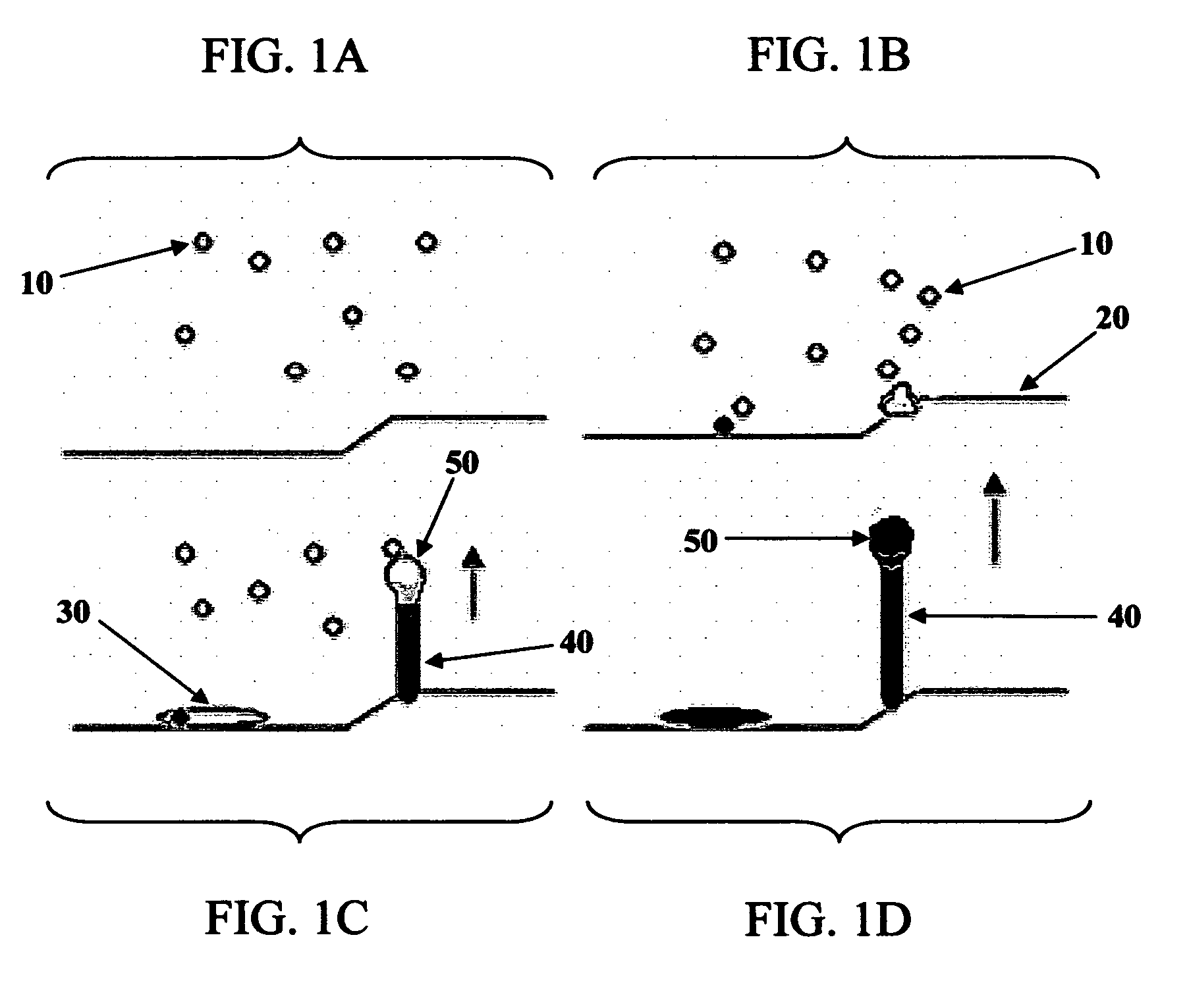
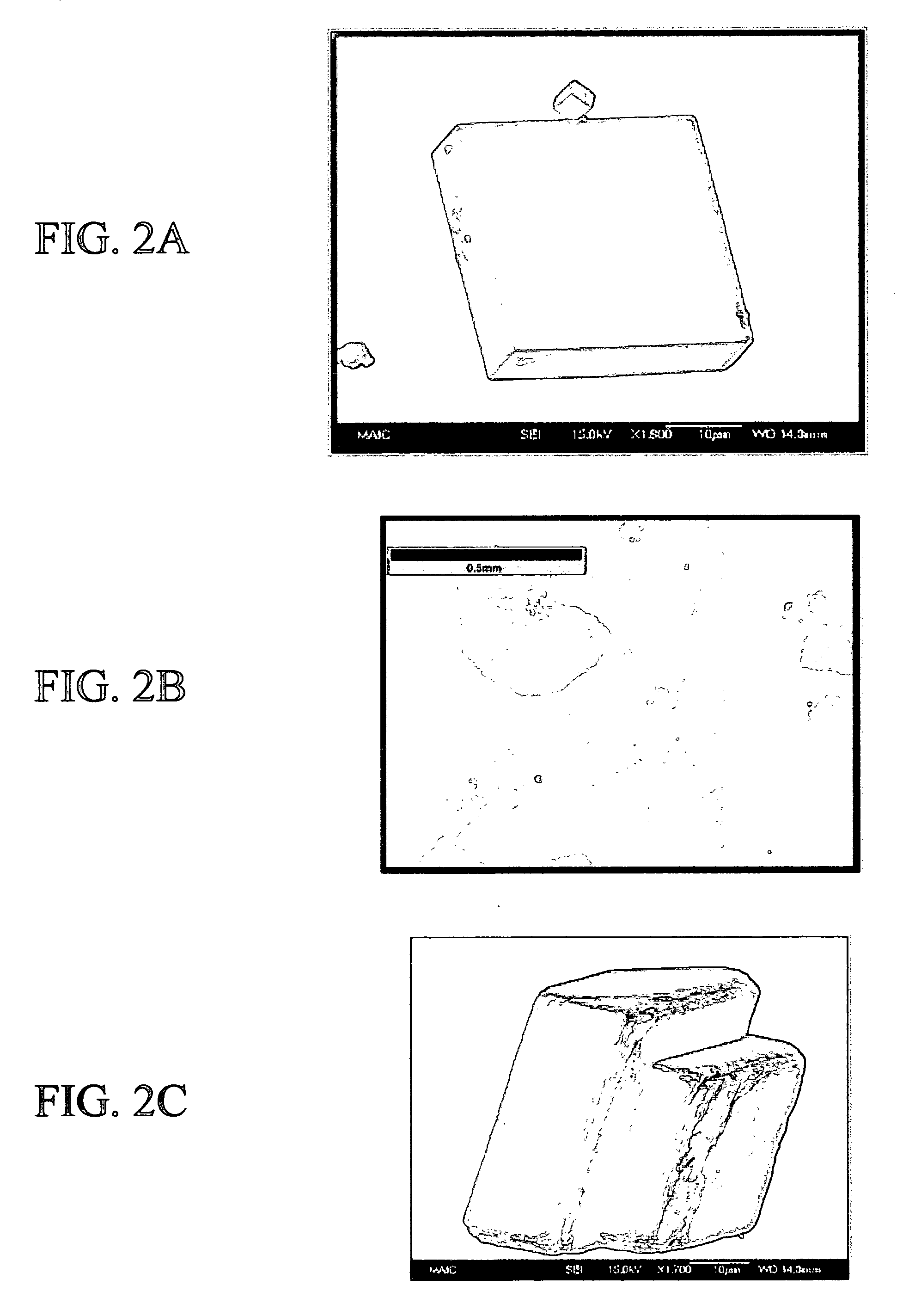
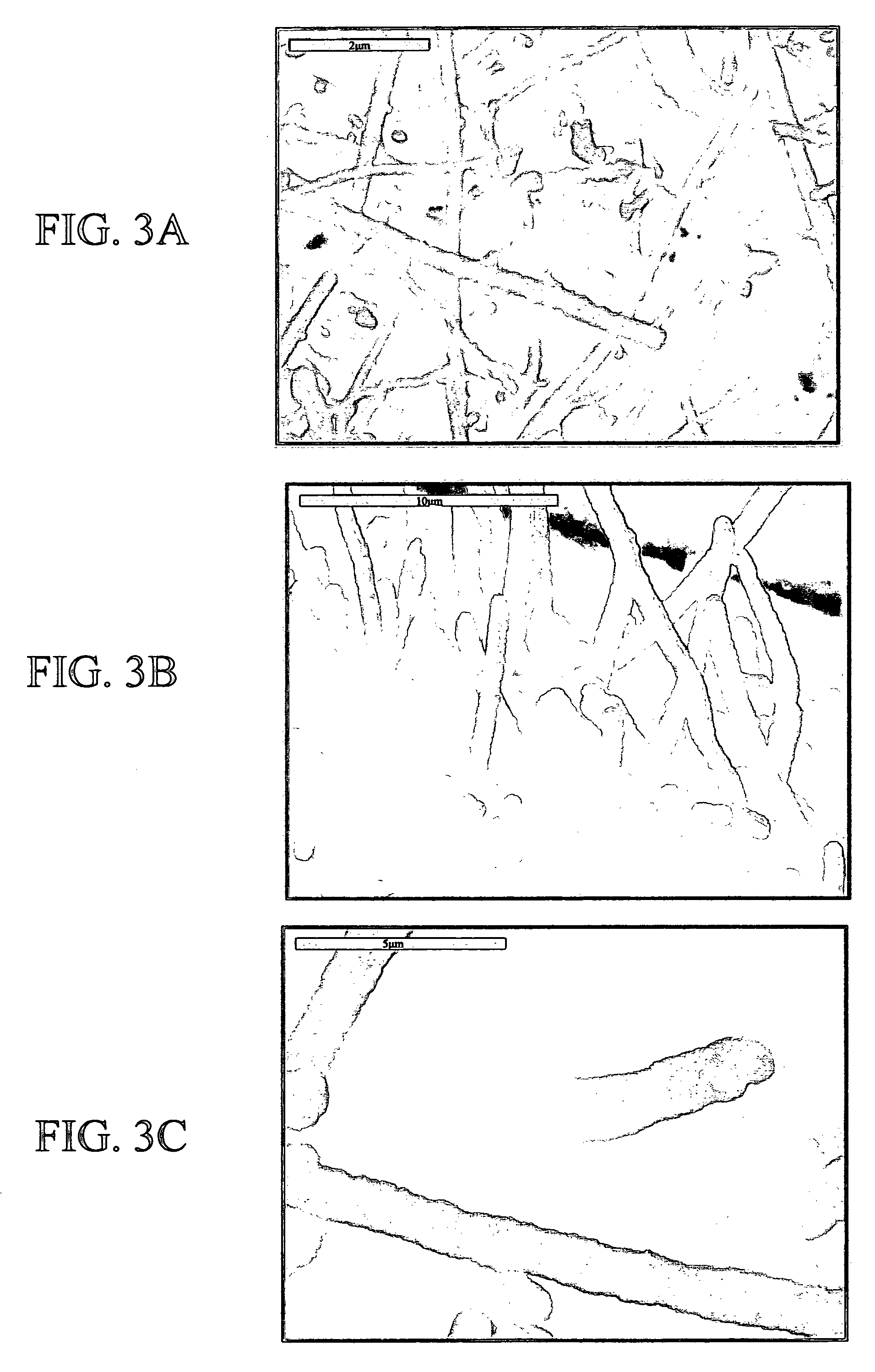
[0063]Fibrous calcium carbonate (CaCO3 / calcite) crystals 100-800 nm in diameter were produced at temperatures as low as 4° C. The calcium carbonate fibers were deposited onto calcite rhombs using a mineralization process involving the inclusion of short-chained acidic polymers (e.g., polyaspartic acid or polyacrylic acid) to a supersaturated calcium carbonate system to induce an amorphous liquid-phase precursor. This latter process, documented in the literature, is known as the polymer-induced liquid-precursor (PILP) process. In previous reports, the present inventors have demonstrated that when the PILP phase is deposited onto an amorphous glass substrate, continuous thin films (˜500 nm thick) of CaCO3 are formed (Gower, L. B. and D. J. Odom [2000] J. Cryst. Grow., 210(4):719-734). Organic substrates, such as type-I collagen, have also been used on which to deposit the PILP phase, resulting in intrafibrillarly mineralized organic / mineral composites (Olszta, ...

example 2

Hydroxyapatite (HA) Fibers

[0067]Enamel, the hardest known vertebrate tissue, is comprised of “rods” of hydroxyapatite tightly packed together in an organized array. Enamel and dentin share a unique starting point, the dentino-enamel junction (DEJ), with the enamel growing away from the DEJ to serve as the mastication surface, and the dentin growing in towards the pulp. While researchers agree that dentine is the first to appear, there is still debate as to whether the enamel epitaxially nucleates from the dentine surface (G. W. Bernard, Ultrastructural observations of initial calcification in dentin and enamel. Journal of Ultrastructure Research 41, 1 (1972).; E. J. Reith, Early stage of amelogenesis as observed in molar teeth of Yorun rats. Journal of Ultrastructure Research 17, 503 (1967).; P. Bodier-Houlle, P. Steuer, J. M. Meyer, L. Bigeard, F. J. G. Cuisinier, High-resolution electron-microscopic study of the relationship between human enamel and dentin crystals at the dentinoe...


PUM

PropertyMeasurementUnit
temperatureaaaaaaaaaa
aspect ratioaaaaaaaaaa

Abstract

The present invention pertains to mineral fibers useful as biomimetics of tissue, such as dental enamel and bone, and as filler in paper, paint, coatings, and plastic production. The present invention also concerns methods for producing mineral fibers by contacting an inorganic liquid-phase mineral precursor with a crystalline inorganic substrate under conditions permitting the inorganic liquid-phase mineral precursor to solidify and crystallize, compositions comprising mineral fibers, and methods for treating a tissue defect within a patient by applying mineral fibers to the site of the tissue defect.

Description

CROSS-REFERENCE TO RELATED APPLICATIONS[0001]This application is a continuation-in-part application of U.S. application Ser. No. 10 / 418,843, filed Apr. 18, 2003, which claims the benefit of U.S. Provisional Application No. 60 / 373,801, filed Apr. 18, 2002; and this application is a continuation-in-part application of U.S. application Ser. No. 10 / 691,002, filed Oct. 22, 2003; which are hereby incorporated by reference in their entirety, including all figures, tables, and drawings.[0002]The subject invention was made with government support under a research project supported by the National Science Foundation Grant No. ECS-9986333. The government has certain rights in this invention.BACKGROUND OF THE INVENTION[0003]Synthetic bone graft material made to closely resemble natural bone would be a useful replacement for natural bone. Acceptable synthetic bone can avoid the problem of availability and harvesting of autogenous bone and the risks and complications associated with allograft bon...

Claims


Application Information

Patent Timeline
Patent Type & Authority Patents(United States)
IPC IPC(8): A61F2/00A61F13/00A61K9/00A61K9/70C12N11/00C12N11/14A61K33/42A61L27/24A61L27/44A61L27/46A61L31/12A61L31/14
CPCA61K33/42A61L27/24A61L27/446A61L27/46A61L31/127A61L31/128A61L31/146B82Y30/00
Inventor GOWER, LAURIE B.OLSZTA, MATTHEW J.
Owner UNIV OF FLORIDA RES FOUNDATION INC
Why Eureka
Try Eureka