
Methods for treating neoplastic disease characterized by vascular endothelial growth factor D expression, for screening for neoplastic disease or metastatic risk, and for maintaining vascularization of tissue

a growth factor d and endothelial growth factor technology, applied in the field of neoplastic disease treatment methods, can solve the problems of tightly controlled and limited angiogenesis, difficult identification of lymphatic vessels, and reduced immune response to tumors.

Inactive Publication Date: 2009-05-19
VEGENICS PTY LTD
View PDF7 Cites 8 Cited by
  • Summary
  • Abstract
  • Description
  • Claims
  • Application Information

AI Technical Summary

Benefits of technology

[0040]According to a first aspect, the present invention provides a method of treating an organism suffering from a neoplastic disease characterized by the expression of VEGF-D by a tumor including, but not limited to, melanomas, breast ductal carcinoma, squamous cell carcinoma, prostate tumors and endometrial cancer. The method comprises screening an organism to determine a presence or an absence of VEGF-D-expressing tumor cells; selecting the organism determined from the screening to have a tumor expressing VEGF-D; and administering an effective amount of a VEGF-D antagonist in the vicinity of said tumor to prevent binding of VEGF-D to its corresponding receptors.
[0057]It will be clearly understood that for the purposes of this specification the term “sample” includes, but is not limited to, obtaining a sample of tissue, blood, serum, plasma, urine, ascities fluid or pleural effusion. Preferably the tissue is human tissue. The compound is preferably a monoclonal antibody. It will be appreciated that use of the second compound helps the practitioner to confirm that the VEGF-D found on the vessels in or near the tumor arises due to receptor-mediated uptake, which supports the hypothesis that VEGF-D, secreted by tumor cells, binds and accumulates in target endothelial cells thereby establishing a paracrine mechanism regulating tumor angiogenesis.
[0061]It will be appreciated that use of the second compound helps the practitioner to confirm that the VEGF-D found on the lymphatic vessels in or near the tumor arises due to receptor- mediated uptake, which supports the hypothesis that VEGF-D, secreted by tumor cells, binds and accumulates in target lymphatic endothelial cells thereby establishing a paracrine mechanism regulating tumor lymphangiogenesis.
[0063]It is contemplated that the fifth aspect is important where VEGF-D / VEGFs are limiting in the tissues of patients, especially in older patients in whom peripheral vessels may be in a state of atrophy. Treatment with an effective amount of VEGF-D could help maintain the integrity of the vasculature by stimulating endothelial cell proliferation in aging / damaged vessels.

Problems solved by technology

In the adult, angiogenesis is tightly controlled and limited under normal circumstances to the female reproductive system.
Recent literature indicates that VEGF blocks maturation of dendritic cells and thereby reduces the effectiveness of the immune response to tumors (many tumors secrete VEGF) (Gabrilovich et al., Blood 92: 4150-4166, 1998; Gabrilovich et al., Clinical Cancer Research 5: 2963-2970, 1999).
Lymphatic vessels are difficult to identify due to the absence of known specific markers available for them.
Disruption of the VEGFR genes results in aberrant development of the vasculature leading to embryonic lethality around midgestation.
However, the role of VEGFR-3 in the development of the lymphatic vasculature could not be studied in these mice because the embryos died before the lymphatic system emerged.
These features are seen in all species infected by orf and can result in the formation of a tumor-like growth or nodule due to viral replication in epidermal cells.
Generally orf virus infections resolve in a few weeks, but severe infections that fail to resolve without surgical intervention are seen in immune impaired individuals.
The localization of VEGF-D protein in human cancer has not been studied due to the lack of antibodies specific for the VHD of VEGF-D.

Method used


Image

  • Methods for treating neoplastic disease characterized by vascular endothelial growth factor D expression, for screening for neoplastic disease or metastatic risk, and for maintaining vascularization of tissue
  • Methods for treating neoplastic disease characterized by vascular endothelial growth factor D expression, for screening for neoplastic disease or metastatic risk, and for maintaining vascularization of tissue
  • Methods for treating neoplastic disease characterized by vascular endothelial growth factor D expression, for screening for neoplastic disease or metastatic risk, and for maintaining vascularization of tissue

Examples


example 1

Production of Monoclonal Antibodies that Bind to Human VEGF-D

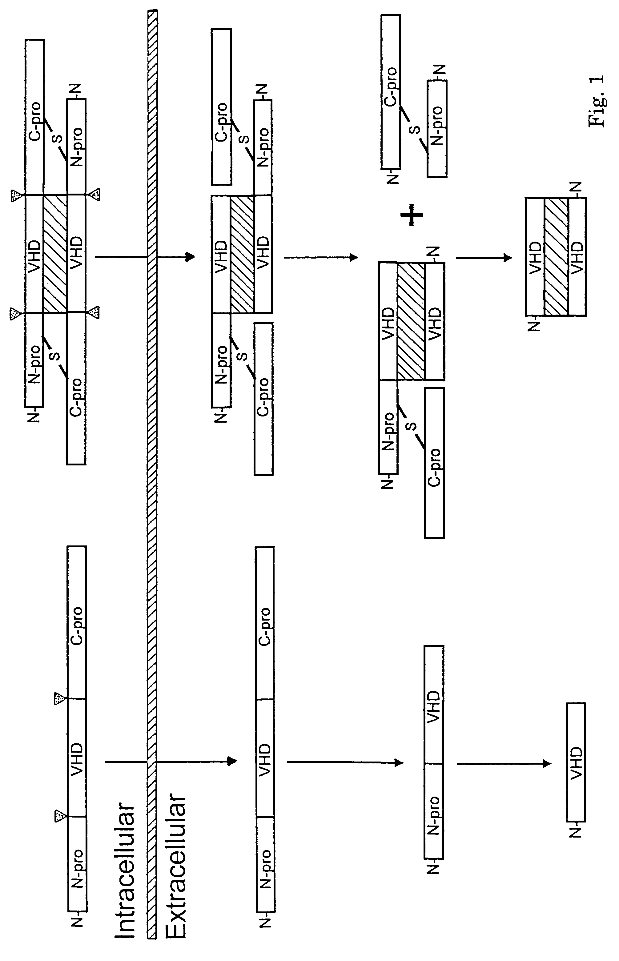
[0113]In order to detect the VEGF Homology Domain (VHD) rather than the N- and C-terminal propeptides, monoclonal antibodies to the mature form of human VEGF-D (residues 93 to 201 of full-length VEGF-D (SEQ ID NO:2), i.e. with the N- and C-terminal regions removed) were raised in mice. A DNA fragment encoding residues 93 to 201 was amplified by polymerase chain reaction (PCR) with Pfu DNA polymerase, using as template a plasmid comprising full-length human VEGF-D cDNA (SEQ ID NO:1).

[0114]The amplified DNA fragment, the correctness of which was confirmed by nucleotide sequencing, was then inserted into the expression vector pEFBOSSFLAG (a gift from Dr. Clare McFarlane at the Walter and Eliza Hall Institute for Medical Research (WEHI), Melbourne, Australia) to give rise to a plasmid designated pEFBOSVEGF-DΔNΔC. The pEFBOSSFLAG vector contains DNA encoding the signal sequence for protein secretion from the interleukin-3 (IL-3...

example 2

Specificity of MAb 4A5

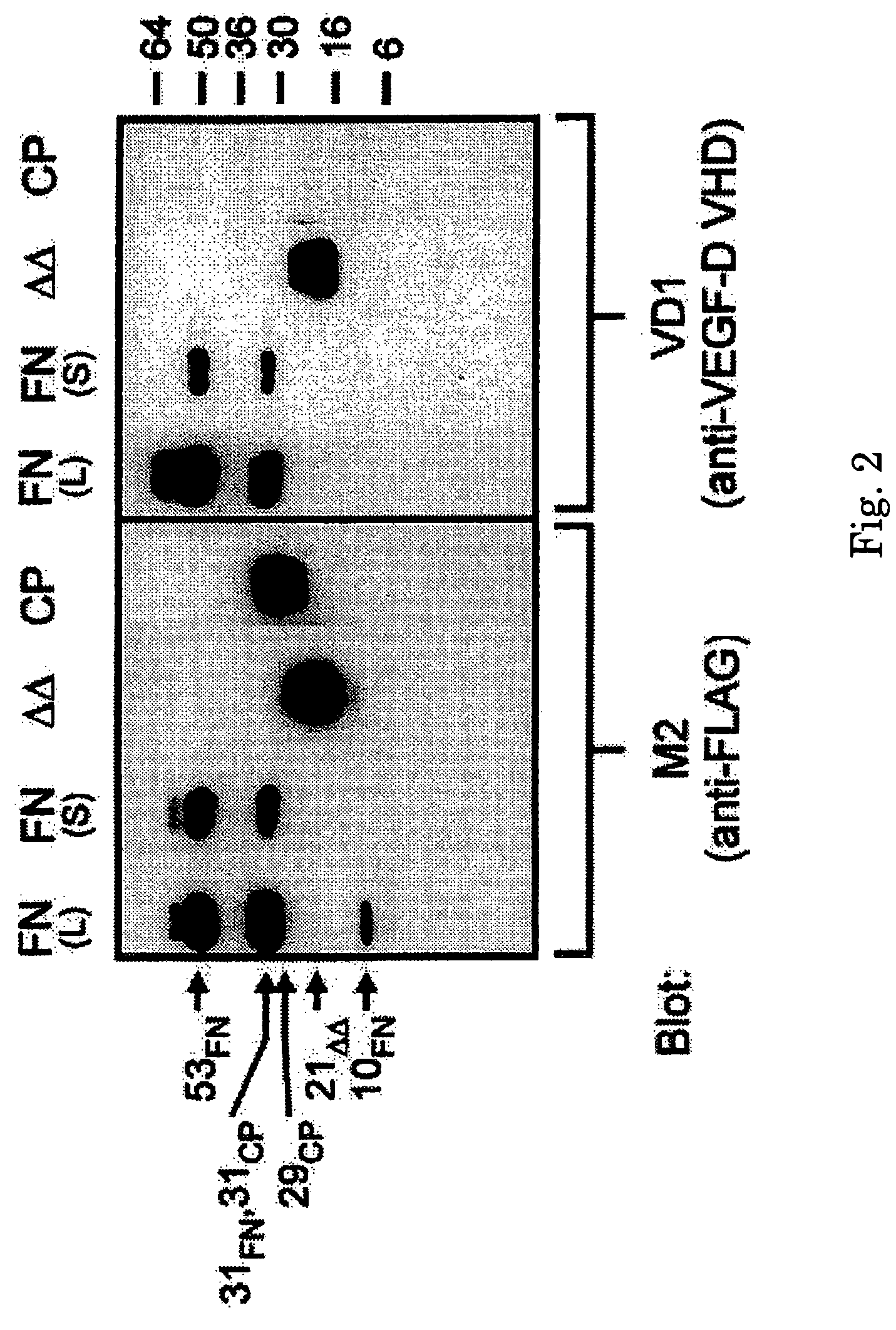
[0120]The specificity of MAb 4A5 (renamed VD1) for the VHD of human VEGF-D was assessed by Western blot analysis. Derivatives of VEGF-D used were VEGF-DΔNΔC, consisting of amino acid residues 93 to 201 of human VEGF-D tagged at the N-terminus with the FLAG® octapeptide (Example 1), VEGF-D-FULL-N-FLAG, consisting of full-length VEGF-D tagged at the N-terminus with FLAG® (Stacker, S. A. et al., J Biol Chem 274: 32127-32136, 1999), and VEGF-D-CPRO, consisting of the C-terminal propeptide, from amino acid residues 206 to 354, which was also tagged with FLAG® at the N-terminus.

[0121]These proteins were expressed in 293-EBNA-1 cells, purified by affinity chromatography with M2 (anti-FLAG®) MAb (IBI / Kodak, New Haven, Conn.) using the procedure set forth in Achen, M. et al., Proc Natl Acad Sci USA 95: 548-553, 1998. Fifty nanograms of purified VEGF-D-FULL-N-FLAG (FN), VEGF-DΔNΔC (ΔΔ), and VEGF-D-CPRO (CP) were analyzed by SDS-PAGE (reducing) and by Western blot using t...

example 3

In Situ Hybridization Studies of VEGF-D Gene Expression in Mouse Embryos

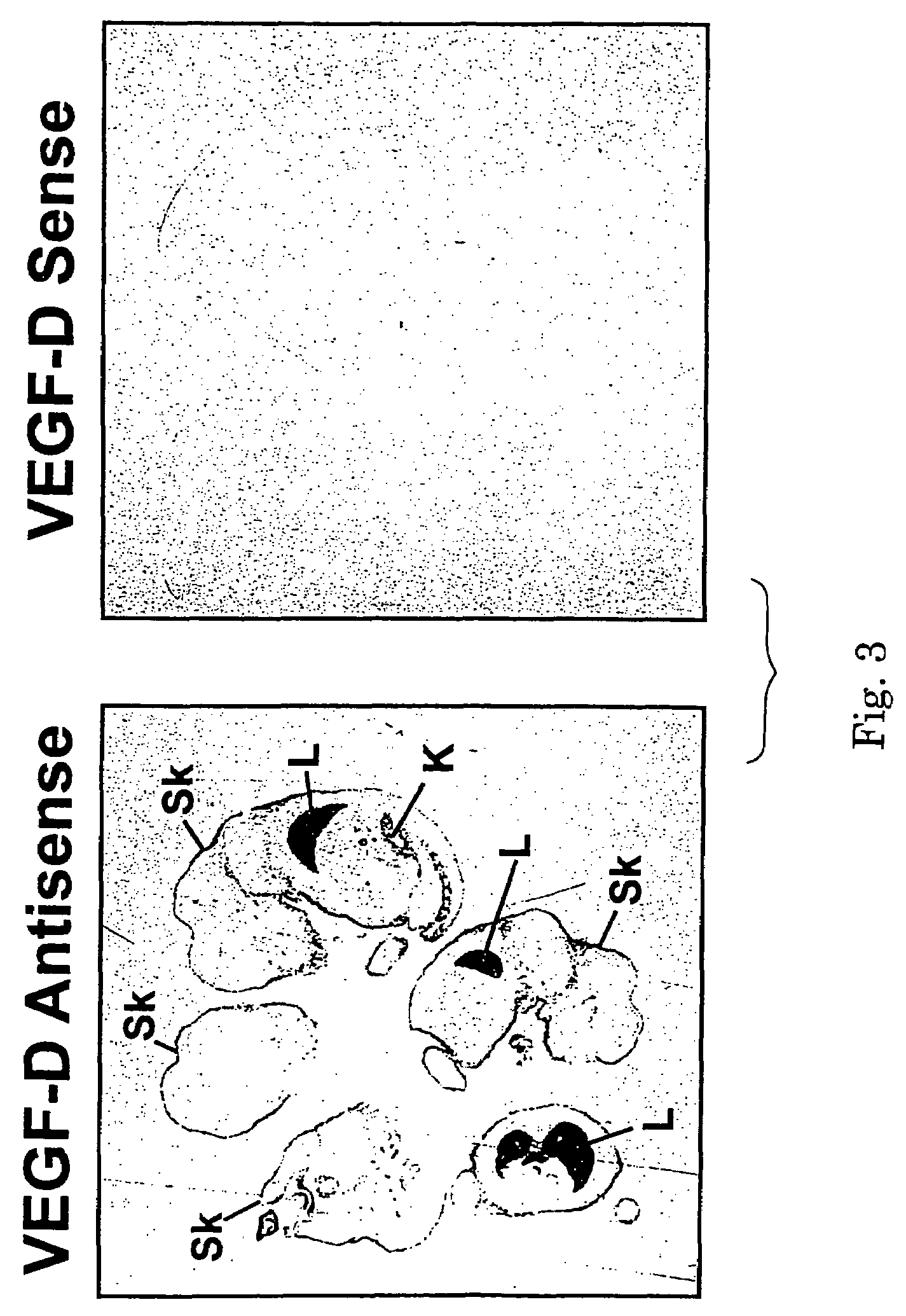
[0125]The pattern of VEGF-D gene expression was studied by in situ hybridization using a radiolabeled antisense RNA probe corresponding to nucleotides 1 to 340 of the mouse VEGF-D1 cDNA (SEQ ID NO:4). The antisense RNA was synthesized by in vitro transcription with T3 RNA polymerase and [35S] UTPαs. Mouse VEGF-D is fully described in International Patent application PCT / US97 / 14696 (WO 98 / 07832). This antisense RNA probe was hybridized to paraffin-embedded tissue sections of mouse embryos at post-coital day 15.5. The labeled sections were subjected to autoradiography for 2 days.

[0126]The resulting autoradiographs for sections hybridized to the antisense RNA and to complementary sense RNA (as negative control) are shown in FIG. 3. In FIG. 3, “L” denotes lung and “Sk” denotes skin, and the two tissue sections shown are serial sections. Strong signals for VEGF-D mRNA were detected in the developing lung and associat...


PUM

PropertyMeasurementUnit
concentrationsaaaaaaaaaa
molecular weightaaaaaaaaaa
vascular permeabilityaaaaaaaaaa

Abstract

A method for treating and alleviating disease characterized by the expression of VEGF-D involving screening to find an organism with tumor cells expressing VEGF-D and administering an effective amount of a VEGF-D antagonist; a method for screening for neoplastic diseas, where detection of VEGF-D on or in a sample such as tumor cells, blood vessel endothelial cells or lymph vessel endothelial cells indicates neoplastic disease; a method for promoting and maintaining vascularization of normal tissue in an organism involving administering a vascularization promoting amount of VEGF-D or a fragment or analog thereof to the organism; a method for screening tumors for metastatic risk involving detecting expression of VEFG-D by a tumor which indicates metastatic risk; and a method of detecting micro-metastasis of neoplastic disease involving detection of VEGF-D on or in a tissue sample which indicates metastasis of a neoplastic disease.

Description

CROSS-REFERENCE TO RELATED APPLICATIONS[0001]This application is a continuation of U.S. application Ser. No. 09 / 956,095, filed Sep. 20, 2001, now abandoned, which is a continuation-in-part of U.S. application Ser. No. 09 / 796,714 filed on Mar. 2, 2001, now abandoned and claims priority of U.S. Provisional Application No. 60 / 186,361, filed Mar. 2, 2000 and U.S. Provisional Application No. 60 / 234,196, filed Sep. 20, 2000.BACKGROUND OF THE INVENTION[0002]The invention relates to a method for treating and alleviating melanomas and various cancers, methods for screening for neoplastic diseases, and a method for promoting and maintaining vascularization of normal tissue.[0003]The two major components of the mammalian vascular system are the endothelial and smooth muscle cells. The endothelial cells form the lining of the inner surface of all blood vessels and lymphatic vessels in the mammal. The formation of new blood vessels can occur by two different processes, vasculogenesis or angiogen...

Claims


Application Information

Patent Timeline
Patent Type & Authority Patents(United States)
IPC IPC(8): G01N33/53C12P21/08G01N33/559G01N33/561G01N33/574G01N33/577A61K38/19C07K16/22C07K16/24
CPCC07K16/22C07K16/24G01N33/57484A61K38/1866A61K2039/505G01N2333/475G01N2333/52
Inventor ACHEN, MARCSTACKER, STEVEN
Owner VEGENICS PTY LTD
Learn More