Method and device for detecting ammonia odors and helicobacter pylori urease infection
a technology of helicobacter pylori and ammonia odor, which is applied in the field of methods and devices can solve the problems of many systems for detecting ammonia odors, expensive instruments, and unsuitable for use by untrained users
- Summary
- Abstract
- Description
- Claims
- Application Information
AI Technical Summary
Benefits of technology
Problems solved by technology
Method used
Image
Examples
example 1
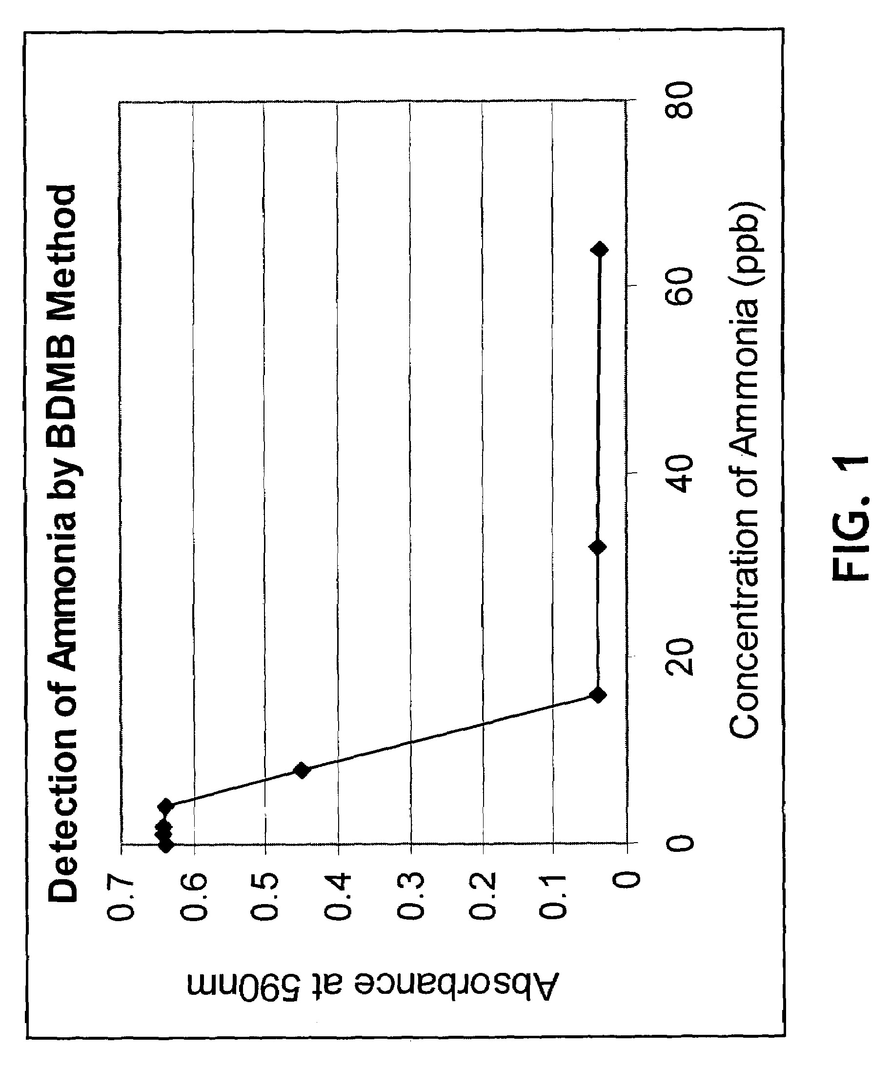
[0058]A reaction mixture was placed into each of 8 vials containing 50 μl of ammonia hydroxide solution as an ammonia source (0, 0.01, 0.02, 0.04, 0.08, 0.16 and 0.64% of ammonia hydroxide, respectively) and 150 μl of MH dye (20 μl of 10.0 mg / ml MH in CH3CN with 5.0 ml of 40 mM sodium acetate and 4M guanidine HCl, pH5.1). After incubation of all the vials at room temperature for less than 4 minutes, a 200 μl portion from each vial was transferred to a microtiter plate well, and the absorbances were measured at 590 nm using a microtiter plate reader (The absorbances can also be measured in the range of 580-615 nm).
[0059]As shown in FIG. 1, a standard curve was derived by plotting the absorbance readings against the concentrations (ppb) of ammonia solutions. In FIG. 1, the x-axis is the concentration of ammonia in parts per billion (ppb) from 10 to 400 and the y-axis is the absorbance at 590 nm from 1 to 0.7. The sensitivity of ammonia detection by MH was shown to be very high.
example 2
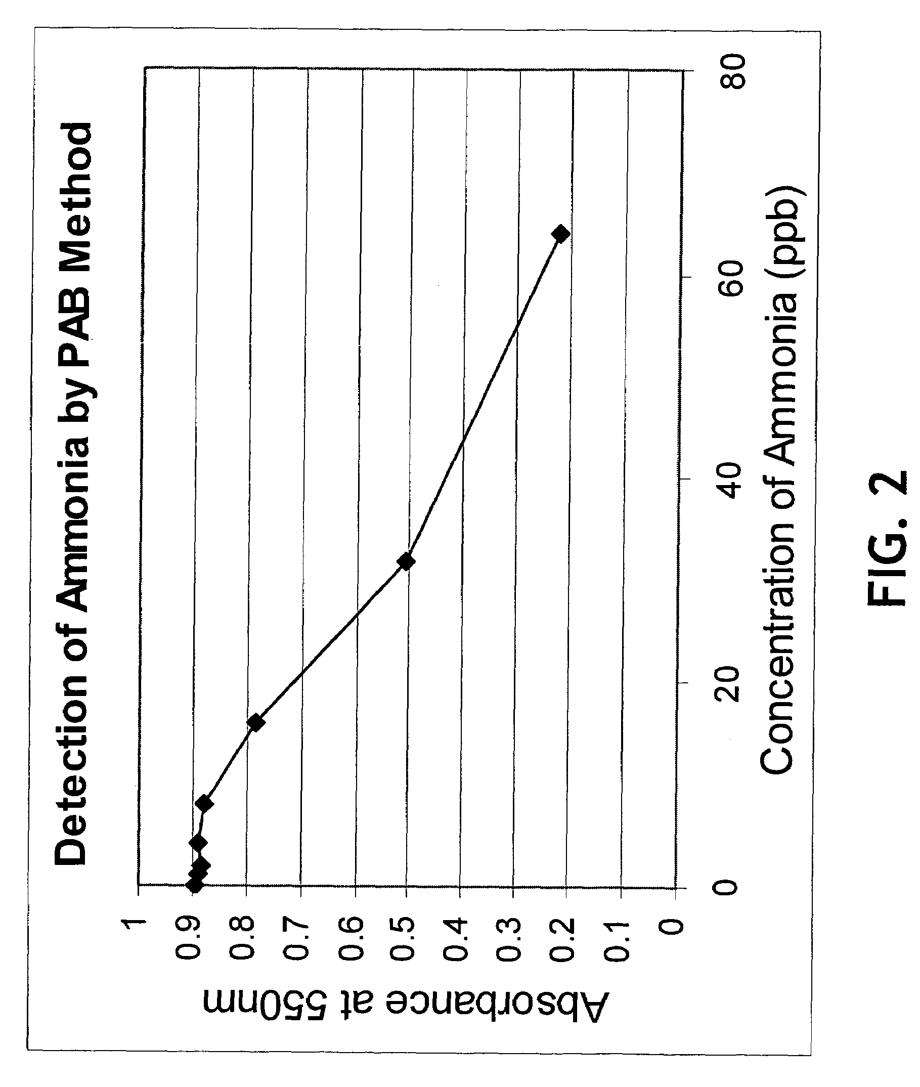
[0060]A similar study was carried out with another dye, pararosaniline base (PAB), which was shown to be sensitive to amine and ammonia odors. In order to generate a standard curve (FIG. 2), a reaction mixture was placed into each of 8 vials containing 50 μl of an ammonia hydroxide solution as an ammonia source (0, 0.01, 0.02, 0.04, 0.08, 0.16 and 0.64% of ammonia hydroxide, respectively) and 150 μl of PAB solution (10 μl of 10 mg / ml PAB stock solution made in CH3CN with 5.0 ml of 40 mM sodium acetate and 4 M guanidine HCl, pH5.1). 200 μl of each reaction mixture was transferred to a mictotiter plate well and the wells were incubated at room temperature for 4 to 5 min. The absorbances were then read at 550 nm using a microplate reader. PAB was shown to be highly selective for ammonia and amine odors. In FIG. 2, the x-axis is the concentration of ammonia in parts per billion (ppb) from 10 to 400 and the y-axis is the absorbance at 550 nm from 1 to 1.0.
example 3
[0061]PAB was then used to see if it was suitable for use in monitoring the reaction in which urease catalyzes urea to ammonia and carbon dioxide by-products (FIG. 3). Into each of two vials (expt. 1 and expt. 2) was placed 1 ml of a reaction mixture containing 100 μl of 10 mM urea, 850 μl of 10 mM PBS, pH7.3, and 50 μl of 10.0 mg / ml urease. Three control vials were prepared, the first control excluding both urea and urease (control 1), the second control excluding urease but containing urea (control 2), and the third control excluding urea but containing urease (control 3). The vials were vortexed and 50 μl from each vial was transferred to a microtiter plate well. PAB solution (10 μl of 10 mg / ml PAB stock solution made in CH3CN with 5.0 ml of 40 mM sodium acetate and 4 M guanidine HCl, pH5.1) was then added to each well and the absorbance change with time was monitored at 550 nm using a microplate reader. In FIG. 3, the x-axis is time in minutes and the y-axis is the absorbance at...
PUM
| Property | Measurement | Unit |
|---|---|---|
| size | aaaaa | aaaaa |
| surface area | aaaaa | aaaaa |
| size | aaaaa | aaaaa |
Abstract
Description
Claims
Application Information
 - R&D
- Intellectual Property
- Life Sciences
- Materials
- Tech Scout
- Unparalleled Data Quality
- Higher Quality Content
- 60% Fewer Hallucinations
Browse by: Latest US Patents, China's latest patents, Technical Efficacy Thesaurus, Application Domain, Technology Topic, Popular Technical Reports.
© 2025 PatSnap. All rights reserved.Legal|Privacy policy|Modern Slavery Act Transparency Statement|Sitemap|About US| Contact US: help@patsnap.com


