Amyloid beta protein (globular assembly and uses thereof)
a technology of globular assembly and myloid beta protein, which is applied in the field of myloid proteins and detection, prevention and treatment of alzheimer's disease, can solve the problems of difficult to distinguish which events are associated with early, causative processes
- Summary
- Abstract
- Description
- Claims
- Application Information
AI Technical Summary
Benefits of technology
Problems solved by technology
Method used
Image
Examples
example 1
Preparation of Amyloid β Oligomers
[0027]1 mg of solid amyloid β1-42 is dissolved in 44 μL of anhydrous DMSO, and this 5 mM solution is diluted into cold (4° C.) F12 media (Gibco) to a total volume of 2.20 mL (50-fold dilution), and vortexed for 30 seconds. The mixture is allowed to incubate at 0° C. for 24 h, followed by centrifugation at 14,000 g for 10 minutes at 4° C. The supernatant is diluted by factors of 1:100 to 1:10,000 into the particular defined medium, prior to incubation with brain slice cultures, cell cultures or binding protein preparations. A 20 μL aliquot of the 1:100 dilution is applied to the surface of a freshly cleaved mica disk, and analyzed by atomic force microscopy. The AFM analysis is carried out on a Digital Instruments Nanoscope IIIa instrument using a J-scanner and operating in tapping mode. AFM data is analyzed using the Nanoscope IIIa software and the IGOR Pro™ waveform analysis software. Analysis by gel electrophoresis was carried out on 15% polyacryl...
example 2
Crosslinking of Amyloid β Oligomers
[0028]Oligomer preparation was carried out as described in example 1, with the following modification. The F12 media was replaced by a buffer containing the following components: N, N-dimethylglycine (766 mg / L), D-glucose (1.802 g / L), calcium chloride (33 mg / L), copper sulfate pentahydrate (25 mg / L), iron(II) sulfate heptahydrate (0.8 mg / L), potassium chloride (223 mg / L), magnesium chloride (57 mg / L), sodium chloride (7.6 g / L), sodium bicarbonate (1.18 g / L), disodium hydrogen phosphate (142 mg / L), and zinc sulfate heptahydrate (0.9 mg / L). The pH of the buffer was adjusted to 7.0 using 0.1 M sodium hydroxide. The supernatant was treated with 0.22 mL of a 25% aqueous solution of glutaraldehyde (Aldrich), followed by 0.67 mL of 0.175 M sodium borohydride in 0.1 NaOH. The mixture stirred at 4° C. for 15 min. and was quenched by addition of 1.67 mL of 20% aqueous sucrose. The mixture was concentrated 5 fold on a SpeedVac and dialyzed to remove component...
example 3
Brain Slice Assay
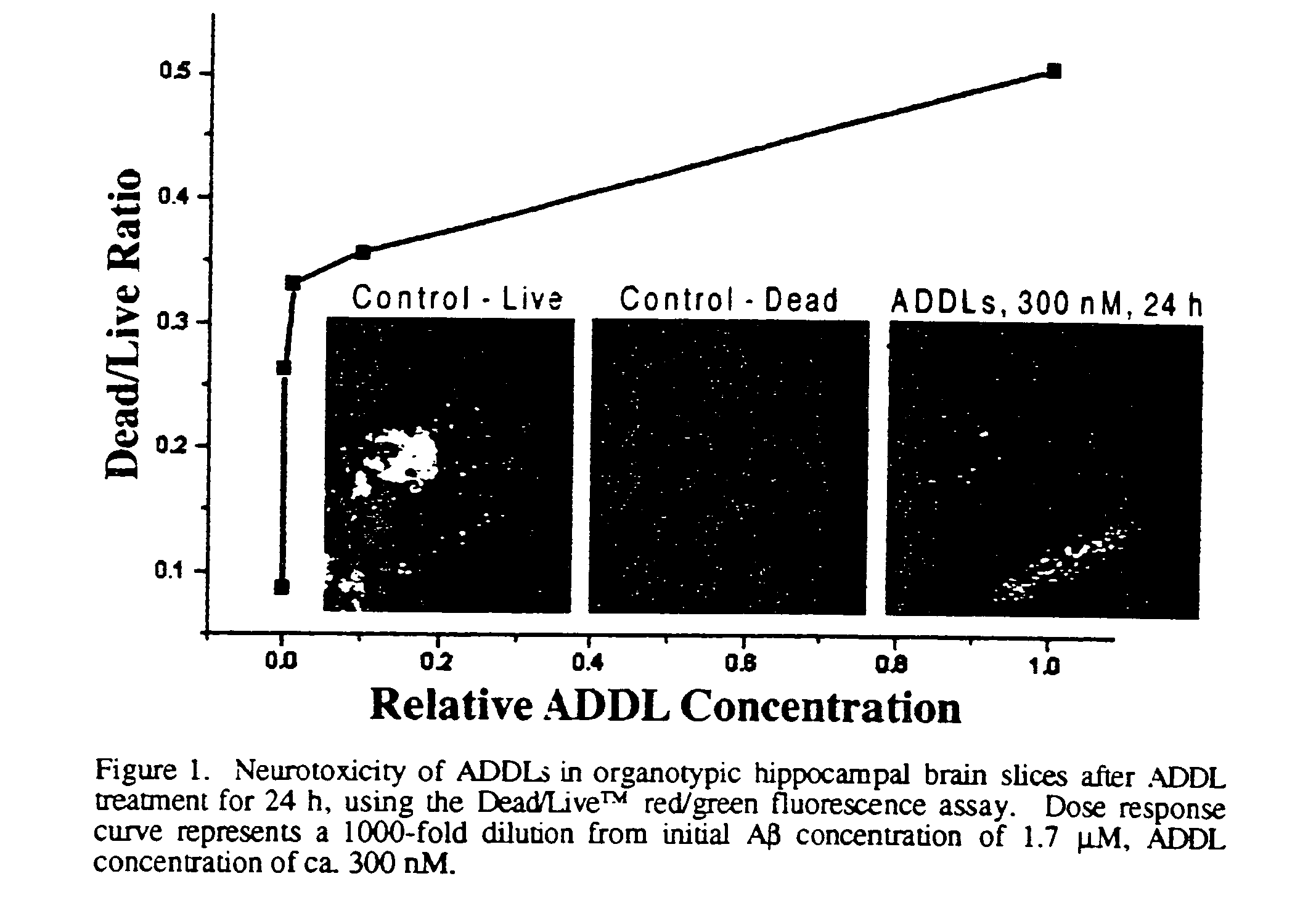
[0029]An adult mouse is sacrificed by carbon dioxide inhalation, followed by rapid decapitation. The head is emersed in cold, sterile dissection buffer (94 ml Gey's balanced salt solution, pH 7.2, supplemented with 2 mL 0.5M MgCl2, 2 ml 25% glucose, and 2 mL 1.0 M Hepes.), after which the brain is removed and placed on a sterile Sylgard-coated plate. The cerebellum is removed and a mid-line cut is made to separate the cerebral hemispheres. Each hemisphere is sliced separately. The hemisphere is placed with the mid-line cut down and a 30 degree slice from the dorsal side is made to orient the hemisphere. The hemisphere is glued cut side down on the plastic stage of a Campden tissue chopper (previously wiped with ethanol) and emersed in ice cold sterile buffer. Slices of 200 μm thickness are made from a lateral to medial direction, collecting those in which the hippocampus is visible. Each slice is transferred with the top end of a sterile pipette to a small petri dis...
PUM
| Property | Measurement | Unit |
|---|---|---|
| total volume | aaaaa | aaaaa |
| flow rate | aaaaa | aaaaa |
| flow rate | aaaaa | aaaaa |
Abstract
Description
Claims
Application Information
 - R&D
- Intellectual Property
- Life Sciences
- Materials
- Tech Scout
- Unparalleled Data Quality
- Higher Quality Content
- 60% Fewer Hallucinations
Browse by: Latest US Patents, China's latest patents, Technical Efficacy Thesaurus, Application Domain, Technology Topic, Popular Technical Reports.
© 2025 PatSnap. All rights reserved.Legal|Privacy policy|Modern Slavery Act Transparency Statement|Sitemap|About US| Contact US: help@patsnap.com


