Method and apparatus for detection of analyte using an acoustic device

a technology of acoustic device and analyte, which is applied in the direction of biological material analysis, biomass after-treatment, synthetic resin layered products, etc., can solve the problems of increasing the risk of breast cancer, increasing the concentration of analyte, and general concentration problems, so as to improve the detection limit of analyte, the effect of low concentration and higher avidity of the sensing surfa

Inactive Publication Date: 2010-01-19
BIOSCALE
View PDF203 Cites 33 Cited by
  • Summary
  • Abstract
  • Description
  • Claims
  • Application Information

AI Technical Summary

Benefits of technology

[0023]The present invention provides improved measurement of analytes. In certain embodiments, the analytes can include small molecules including but not limited to estradiol and therapeutic drugs. As a result of the present invention, the pharmacokinetics of a given drug can be readily determined and drug regimens can be more readily customized (by, for example, a physician) to maintain efficacy of the drug while reducing unwanted side effects. In addition, the present invention can be used at the point of care to measure biologically and / or clinically relevant analytes while avoiding delays (associated with, for example, sending samples to an off-site testing facility) in order to customize care and increase the level of patient compliance. As used herein, the term point of care can include, for example, the doctor's office, clinic, emergency room, and mobile treatment facility (e.g., an ambulance). In one embodiment, the method further comprises adjusting dosage of said therapeutic drug administered to an individual based on the level of said drug in the sample. The level of the therapeutic drug can be varied based on the known therapeutic range for the therapeutic drug being tested.
[0025]The efficiency and low operating cost associated with the system 100 of FIG. 1 is well-suited for running individual samples in a manner that does not require operating in a batch mode where many samples are acquired prior to running an assay. In this manner, embodiments of the present invention are well suited for analyzing samples on an individual basis.
[0027]In one embodiment, the sensing device is a flexural plate wave (FPW) device, which functions particularly well with the magnetic particles for two reasons. First, the presence of the magnetic particles on the surface of the sensing device results in an amplified FPW signal response. The larger combined size and density of the analyte-particle complex yields a larger FPW signal response than the analyte alone. Second, the surface of the sensor in the FPW device consists of a thin membrane that is typically only a few micrometers thick, which allows larger magnetic fields and field gradients to be created at the sensor surface because the field source can be positioned closer to the sample flow. This results in higher fractional capture of the analyte from the sample. With this higher capture rate and efficiency, it is possible to process larger sample volumes in shorter times than would be otherwise possible.
[0032]As a result of the present invention, much lower levels of analyte can be detected compared to conventional assays. The detection limit of analyte is improved by a number of factors. In one embodiment, the present invention uses fewer particles per sample, thereby resulting in particles that are more densely coated with analyte, even where the concentration of analyte in the sample is low. As a result of the presence of higher levels of analyte per particle, each coated particle has a higher avidity for the sensing surface.
[0033]Where the assay format is a competition assay, that is, the sensing surface is coated with the analyte, particles that are more densely coated with analyte are more effectively blocked from binding to the sensing surface because each particle comprises fewer capture agent molecules that are free to bind to the analyte on the sensing surface. In other words, a lower particle to analyte ratio results in better competition between the analyte in the sample and the analyte on the sensing surface which allows for the detection of lower concentrations of analyte in the sample. Thus, the present invention improves the accuracy and the sensitivity of the system because the particles are more densely coated with analyte, and have a higher binding strength for the sensing surface.
[0034]In another embodiment, devices fabricated and methods performed according to the principles of the present invention are particularly useful for capturing low concentrations of particles because the sensor surface is thin (for example, in one embodiment, on the order of several microns). In one embodiment, the thin surface is used in conjunction with a magnetic field gradient and magnetic particles. Because the sensor surface is thin, a large magnetic field gradient can be induced one the side of the sensor surface. The large magnetic field gradient can enhance the attraction of the particles to the sensor surface by focusing the field close to the surface of the sensor, while allowing other material contained in the sample to flow by. The large magnetic field gradient can enhance the attraction of the magnetic particles to the sensor surface by focusing the field close to the surface of the sensor, while allowing other material contained in the sample to flow by. In this way, lower numbers of particles can be used, thereby improving the detection limit of the system, because the magnetic field gradient serves to concentrate the particles near the sensing surface where they can interact with the bound analyte or capture agent, depending on the assay format. In various embodiments, the field gradient can be removed to allow any non-specifically bound particles to be washed away. In various embodiments, the magnetic field gradient can be removed to allow any non-specifically bound particles to be washed away.

Problems solved by technology

Significant challenges for a system that detects analytes (e.g., chemical and biological agents) in liquid media include concentration of the analyte in the media, and transport of the analyte to a sensor surface.
For biological applications, concentration issues generally arise since the concentrations of such analytes tend to be low.
Additionally, biological analytes (e.g., cells, cell fragments and macromolecules such as proteins and nucleic acids) tend to be relatively large; hence, transport issues arise because these larger analytes diffuse in fluid solution very slowly.
An elevated level of estradiol is correlated with increased risk of developing breast cancer.
However, a low estradiol level is correlated with an increased risk for vertebral fractures.
However, tamoxifen can lower serum estradiol levels to a point that increases the incidence of vertebral fracture.
The detection of small molecules such as estradiol is often difficult not only because of potentially low levels in the sample, but also because of the limited number of suitable capture agents that can be used to bind the small molecule analyte.
As a result of such limitations, it is often difficult to design or modify existing assays, such as enzyme-linked immunosorbant assays that are sensitive and specific enough to detect low levels of the small molecule analyte.
The delay associated with analyzing a sample makes it difficult for a doctor to accurately specify a proper treatment.
Acoustic excitation has been used to draw cells to field nodes, but it is difficult to use this technique alone to transport material to a surface.
Electrical fields (electrophoresis and dielectrophoresis) have been used to enhance transport but are not universally applicable to all analytes and sample types.
Furthermore, the electrical properties of microbes can vary within a given species and strain, making it hard to predict system performance under all intended operating conditions.
This requirement can conflict with the optimum binding or wash conditions in an assay.
Also, electrical fields can dissipate energy and heat conductive fluids (e.g., 0.1 M phosphate buffer solution), which is undesirable since heating can damage the biological analytes.

Method used


Image

  • Method and apparatus for detection of analyte using an acoustic device
  • Method and apparatus for detection of analyte using an acoustic device
  • Method and apparatus for detection of analyte using an acoustic device

Examples


example i

Generalized Method for Capture Agent Functionalization of a Surface of a Flexural Plate Wave Device

[0157]1. Deposit gold onto the surface (e.g., sensor surface 143) of the flexural plate wave device 104 and clean the gold surface 143 with, for example, oxygen plasma.

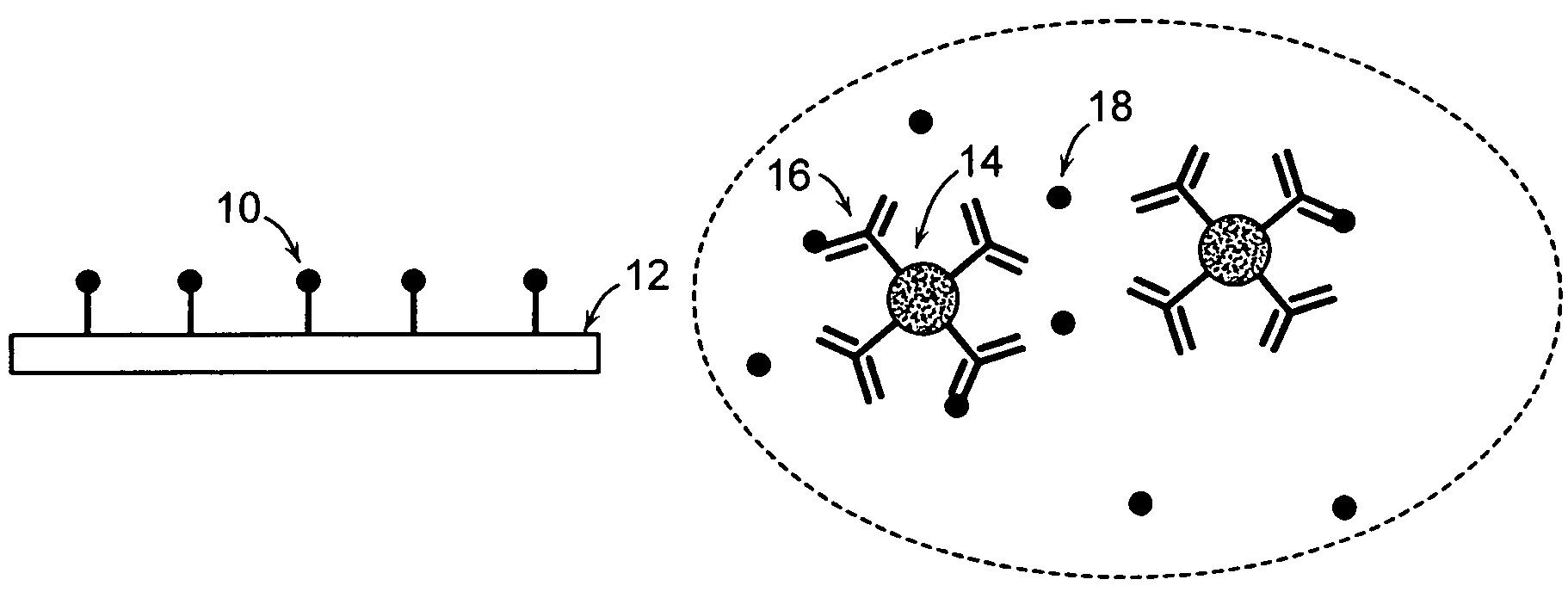
[0158]2. An ideal surface chemistry for the surface 143 of the gold is one that provides 1) non-specific binding resistance and 2) reactive groups located on the surface for covalent attachment of capture agents. An exemplary surface chemistry for the surface 143 of the gold is a self-assembled monolayer (SAM) of alkane thiols. The SAM can be formed from a mixture of two alkane thiols; one terminated with a reactive group for subsequent covalent attachment of capture agents, and one terminated with a non-reactive group. By way of example, a mixture of EG3-OH (EG3) and EG6-OCH2COOH (EG6) terminated C11-alkane thiols may be used for this purpose. In one embodiment, the flexural plate wave device 104 (particularly the surfa...

example ii

Detecting E. coli 0157:H7 In Ground Beef Using, for Example, the Method of FIG. 3

FIG. 5 Contains Data Representative of Various Concentrations of E. coli

[0160]1. Prepare an analyte sample containing E. coli O157:H7 with a concentration greater than about 100 cfu / mL.

[0161]2. Concentrate the analyte sample in solution by performing an immunomagnetic separation. A variety of commercial instruments (e.g., PATHATRIX™ (antibody coated paramagnetic particles) by Matrix Microsciences and BEAD RETRIEVER™ (magnetic bead processor ) by Dynal Biotech) or manual methods may be used to perform the immunomagnetic separation. An exemplary manual method involves:

[0162]a. Resuspend magnetic beads coated with E. coli antibody (e.g., DYNABEADS™ anti-E. coli 0157, available from Dynal Biotech) until the magnetic bead pellet in the bottom of the tube disappears. Place a microcentrifuge tube in the rack (e.g., a Dynal MPC-S) of a magnetic plate. Pipette 1-20 μL of magnetic bead stock solution into the tu...

example iii

Detecting Prostate Specific Antigen (PSA) in Human Blood Serum Using, for Example, the Method Steps of FIG. 3

[0181]1. Prepare an analyte sample containing human serum obtained by centrifugation from a human blood sample.

[0182]2. Concentrate the analyte sample in solution by performing an immunomagnetic separation. A variety of commercial instruments (e.g., PATHATRIX™ (antibody coated paramagnetic particles) by Matrix Microsciences and BEAD RETRIEVER™ (magnetic bead processor) by Dynal Biotech) or manual methods may be used to perform the immunomagnetic separation. An exemplary manual method involves:

[0183]a. Resuspend magnetic beads coated with PSA antibody (e.g., DYNABEADS® anti-PSA, available from Dynal Biotech) until the magnetic bead pellet in the bottom of the tube disappears. Place a microcentrifuge tube in the rack (e.g., a Dynal MPC-S) of a magnetic plate. Pipette 1-20 μL of magnetic bead stock solution into the tube (the volume of magnetic bead stock selected is based on de...


PUM

PropertyMeasurementUnit
volumeaaaaaaaaaa
volumeaaaaaaaaaa
volumeaaaaaaaaaa

Abstract

Methods for detecting analytes in a sample are provided. A plurality of particles, each of which is coated with a capture agent having an affinity for the analyte, is combined with the sample to form a plurality of analyte-particle complexes. The system also includes a transport arrangement for transporting the sample to the sensor surface, and a magnetic field inducing structure constructed and arranged to establish a magnetic field at and adjacent to the sensor surface. The resonant sensor produces a signal corresponding to an amount of analyte-particle complexes that are bound to the sensor surface.

Description

CROSS REFERENCE TO RELATED APPLICATIONS[0001]This application is a continuation-in-part of U.S. Ser. No. 11 / 183,484 filed on Jul. 18, 2005, which claims the benefit of U.S. Provisional Patent Application Ser. No. 60 / 690,592, filed Jun. 15, 2005. This application also claims the benefit of U.S. Provisional Patent Application Ser. No. 60 / 676,759, filed on May 2, 2005, entitled “Methods and Apparatus for Viral Load Detection and Measurement.”TECHNICAL FIELD[0002]The present invention relates to methods for detecting one or more analytes in fluid samples.BACKGROUND OF THE INVENTION[0003]Significant challenges for a system that detects analytes (e.g., chemical and biological agents) in liquid media include concentration of the analyte in the media, and transport of the analyte to a sensor surface. For biological applications, concentration issues generally arise since the concentrations of such analytes tend to be low. Additionally, biological analytes (e.g., cells, cell fragments and ma...

Claims


Application Information

Patent Timeline
Patent Type & Authority Patents(United States)
IPC IPC(8): G01N33/553B32B27/00B32B27/12
CPCB01L3/502761G01N29/022G01N29/036G01N29/4418G01N33/54313G01N33/9493G01N2800/324B01L2300/0681B01L2300/1822B01L2400/043B01L2400/0487B82Y15/00B82Y30/00G01N2291/0256G01N2291/0427B01L2200/0647
Inventor SRIVASTAVA, ALOKWANG, WAYNE U.MILLER, MICHAELMASTERS, BRETT P.LUNDSTROM, MARK
Owner BIOSCALE
Why Eureka
Try Eureka