
Implantable wireless sensor

a wireless sensor and wireless sensor technology, applied in the field of implantable wireless sensors, can solve the problems of high patient injury risk, leakage, and sudden death of aortic arteries, and achieve the effects of reducing the risk of aortic arteries, and improving the safety of patients

Inactive Publication Date: 2010-04-20
ST JUDE MEDICAL LUXEMBOURG HLDG II S A R L SJM LUX II
View PDF138 Cites 63 Cited by
  • Summary
  • Abstract
  • Description
  • Claims
  • Application Information

AI Technical Summary

Benefits of technology

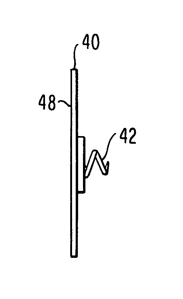
[0012]The present invention comprises a device that can be implanted into the human body using non-surgical techniques to measure a corporeal parameter such as pressure, temperature, or both. Specific target locations could include the interior of an abdominal aneurysm or a chamber of the heart. This sensor is fabricated using MicroElectroMechanical Systems (MEMS) technology, which allows the creation of a flexible device that is small, accurate, precise, durable, robust, biocompatible, radiopaque and insensitive to changes in body chemistry, biology or external pressure. This device will not require the use of wires to relay pressure information externally nor need an internal power supply to perform its function.
[0013]The MEMS approach to sensor design lends itself to the fabrication of small, flat flexible sensors that can be formed using biocompatible polymers as substrate materials. The pressure sensor described above can then be manipulated into a smaller shape and size by rolling, bending, or folding it into a cylindrical form. This smaller object can be introduced into the sac of an abdominal aneurysm at the time an endograft is deployed within the aorta by using standard endovascular catheter techniques. Once inserted in the abdominal aneurysm sac or in a chamber of the heart, the device either on its own or through the addition or inclusion of metallic elements fabricated from stainless steel or super-elastic or shape memory nitinol alloys can unfurl into a preferred flat shape. The metallic components may also include anchors, hooks, harpoons, coils, barbs or other shapes and configurations designed to secure the pressure sensor to the wall on the aneurysm sac or a chamber of the heart. In addition, appropriately biocompatible coatings may be applied to the surface of the sensor to prevent adhesion of biological substances or coagulated blood to the sensor that could interfere with its proper function.
[0016]In a further alternative delivery procedure the sensor can be loaded into the annular space between the inner and outer catheters by inserting the sensor into a longitudinal slit cut into the outer catheter and attaching a tab on the sensor's surface into a slot cut into the inner coaxial catheter. By rotation of the inner tube, the sensor will be retracted through the slit and positioned in the annular space between the two tubes. To deploy the device, the rotation of the inner tube is reversed and the sensor emerges through the slit of the outer catheter. There are two specific advantages to this deployment mechanism. First, the sensor can be packaged and stored in a flat configuration. This is desirable since long term storage in a pre-loaded curved geometry could make it more difficult for the sensor to re-establish the flat arrangement that is optimal for effective electromagnetic inductive coupling with the external read-out unit. The second advantage is that by cutting the longitudinal slit at angle that is offset from the main axis of the outer tube, the sensor will be biased into a planar configuration as it is forced through the slit during the deployment process.

Problems solved by technology

Abdominal aortic aneurysms represent a dilatation and weakening of the abdominal aorta which can lead to aortic rupture and sudden death.
Previously, the medical treatment of abdominal aortic aneurysms required complicated surgery with an associated high risk of injury to the patient.
A significant problem with endografts is that, due to inadequate sealing of the graft with the aorta, leaks can develop that allow blood to continue to fill the aneurysmal sac.
Left undiscovered, the sac will continue to expand and potentially rupture.
These procedures are expensive, not sensitive, and painful.
In addition, they subject the patient to additional risk of injury.
While some of the above objectives are accomplished, this device has multiple problems that would make its use impractical.
Elements of this kind cannot be practically manufactured in dimensions that would allow for endovascular introduction.
In addition, this type of pressure sensor would be subject to many problems in use that would limit its accuracy and reliability.
One example would be exposure of the mechanical sensing element to body fluids which could disrupt its function.
Also, by failing to take advantage of specific approaches to electronic component fabrication, the Van Bockel device requires a complex system for acquiring data from the sensor necessary for the physician to make an accurate determination of intra-aneurysmal pressure.
The Van Bockel device would inherently not be as flexible as required for certain endovascular or percutaneous procedures.

Method used


Image

  • Implantable wireless sensor
  • Implantable wireless sensor
  • Implantable wireless sensor

Examples


Embodiment Construction

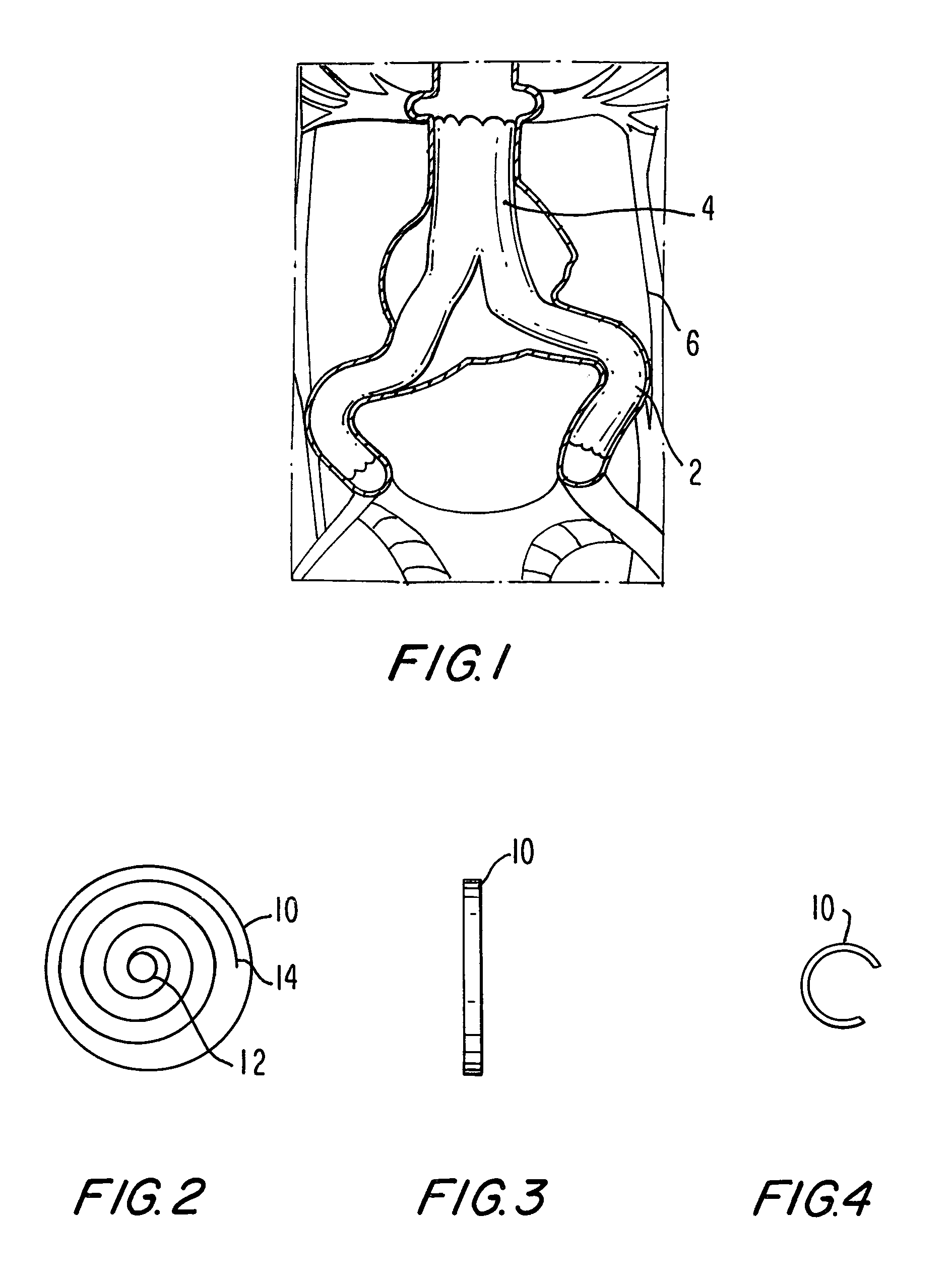
[0050]The invention can perhaps be better understood by referring to the drawings. FIG. 1 represents a typical abdominal aortic aneurysm stent 2 that has been inserted into an abdominal aorta 4. Stent 2, which typically comprises a polymeric material, creates passageways within an aneurysm sac 6.

[0051]One embodiment of a sensor according to the invention is shown in FIGS. 2, 3, and 4, where a disc-shaped sensor 10 comprises a capacitor disk 12 and a wire spiral 14. FIG. 3 is a lateral view of sensor 10, and FIG. 4 is a lateral view of sensor 10 in a folded configuration for insertion. The fact that sensor 10 is sufficiently flexible to be folded as shown in FIG. 4 is an important aspect of the invention.

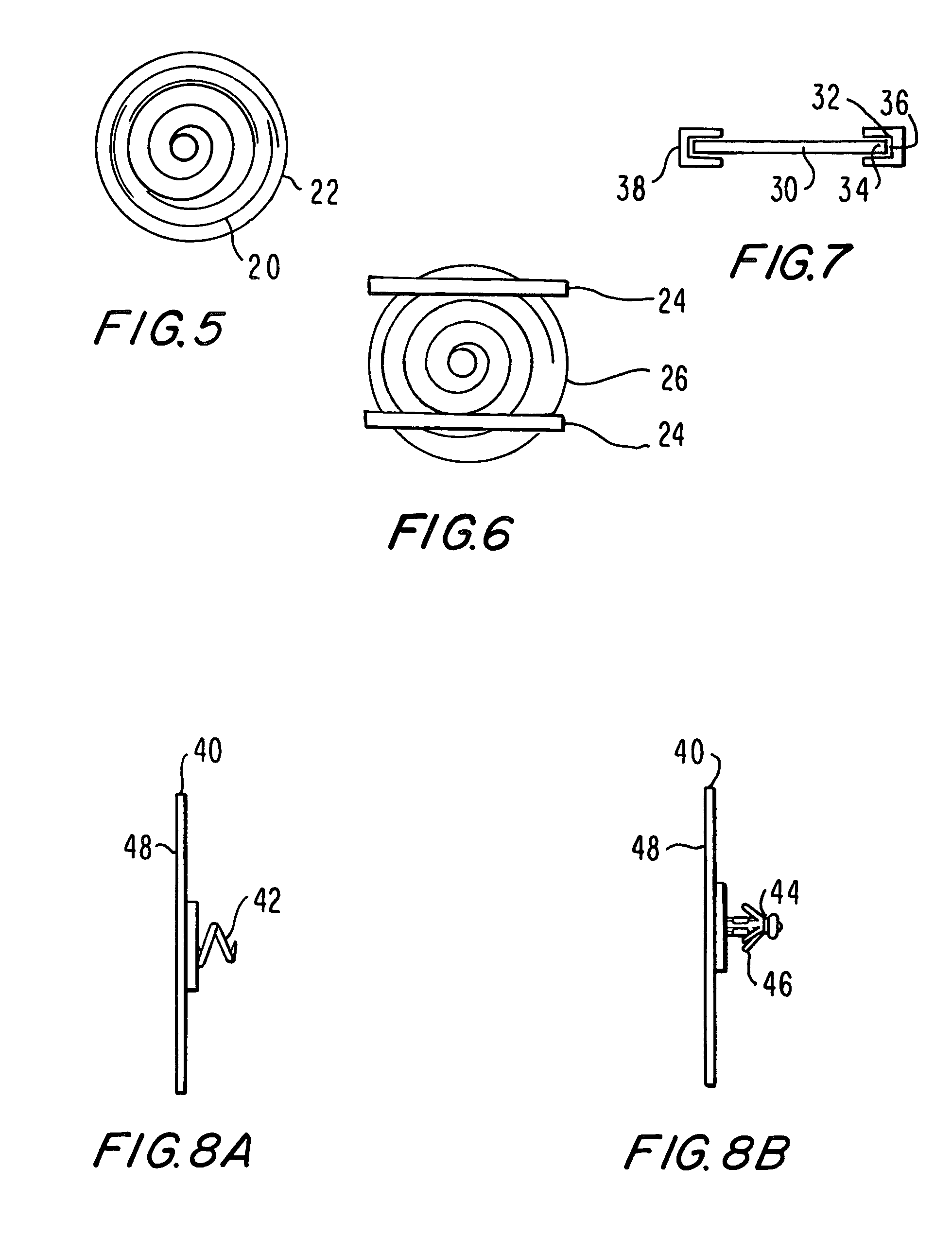
[0052]In FIG. 5 a ring 20 comprised of a shape memory alloy such as nitinol has been attached to, for example, with adhesive, or incorporated into, for example, layered within, a sensor 22, and in FIG. 6 strips 24 comprised of a shape memory alloy such as nitinol have been attached t...


PUM


Abstract

The progress of a endovascular aneurysm repair can be monitored by inserting a pressure transducer sensor using a catheter into the sac during endovascular aneurysm repair and then using a small, hand-held read out device to measure pressure easily, safely, inexpensively and accurately. In one aspect a sensor is introduced into the body by the steps of folding or rolling the sensor into a cylinder, loading it into a catheter, and deploying into the aneurysm sac by allowing it to unroll or unfold, either by itself or facilitated by the incorporation of a super-elastic alloy component.

Description

FIELD OF THE INVENTION[0001]The application is directed to an implantable wireless sensor. More particularly, this invention is directed to a wireless, unpowered, micromechanical, flexible sensor that can be delivered using endovascular techniques, to measure a corporeal parameter such as pressure or temperature.BACKGROUND OF THE INVENTION[0002]Abdominal aortic aneurysms represent a dilatation and weakening of the abdominal aorta which can lead to aortic rupture and sudden death. Previously, the medical treatment of abdominal aortic aneurysms required complicated surgery with an associated high risk of injury to the patient. More recently, endografts (combining stents and grafts into a single device) have been developed that can be inserted through small incisions in the groin. Once in place, these endografts seal off the weakened section of the aorta. The aneurysms can then heal, eliminating the risk of sudden rupture. This less invasive form of treatment for abdominal aortic aneur...

Claims


Application Information

Patent Timeline
IPC IPC(8): A61F13/00A61B5/02A61B5/00A61B5/03
CPCA61B5/03A61B5/0002
Inventor FONSECA, MICHAELALLEN, MARKSTERN, DAVIDWHITE, JASONKROH, JASON
Owner ST JUDE MEDICAL LUXEMBOURG HLDG II S A R L SJM LUX II
Learn More