Agents for magnetic imaging method

a magnetic imaging and agent technology, applied in the field of nuclear magnetic resonance technique, can solve the problems of difficult differentiation of the tissue of interest from the surrounding tissue in the resulting image, low efficiency of magnetic imaging, and high toxicity of metal ions, and achieve the effect of easy internalisation into the cell

Inactive Publication Date: 2010-08-24
BRACCO IMAGINIG SPA
View PDF31 Cites 2 Cited by
  • Summary
  • Abstract
  • Description
  • Claims
  • Application Information

AI Technical Summary

Benefits of technology

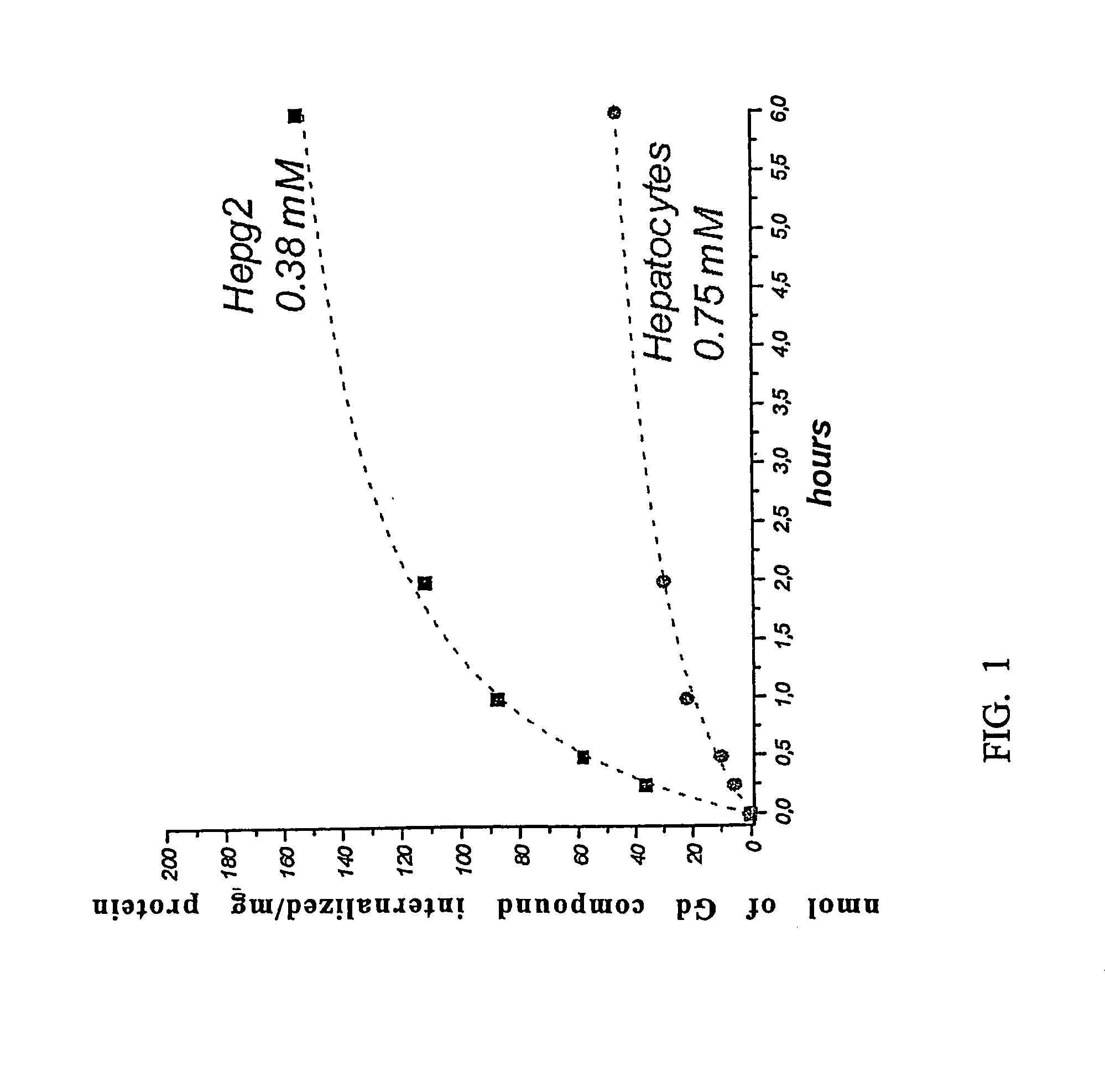
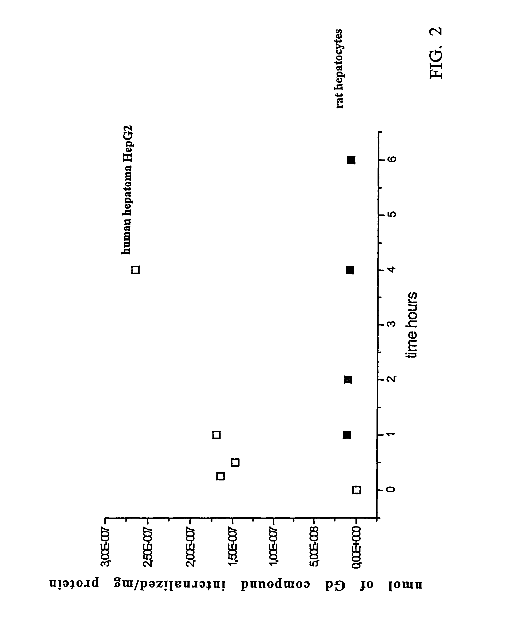
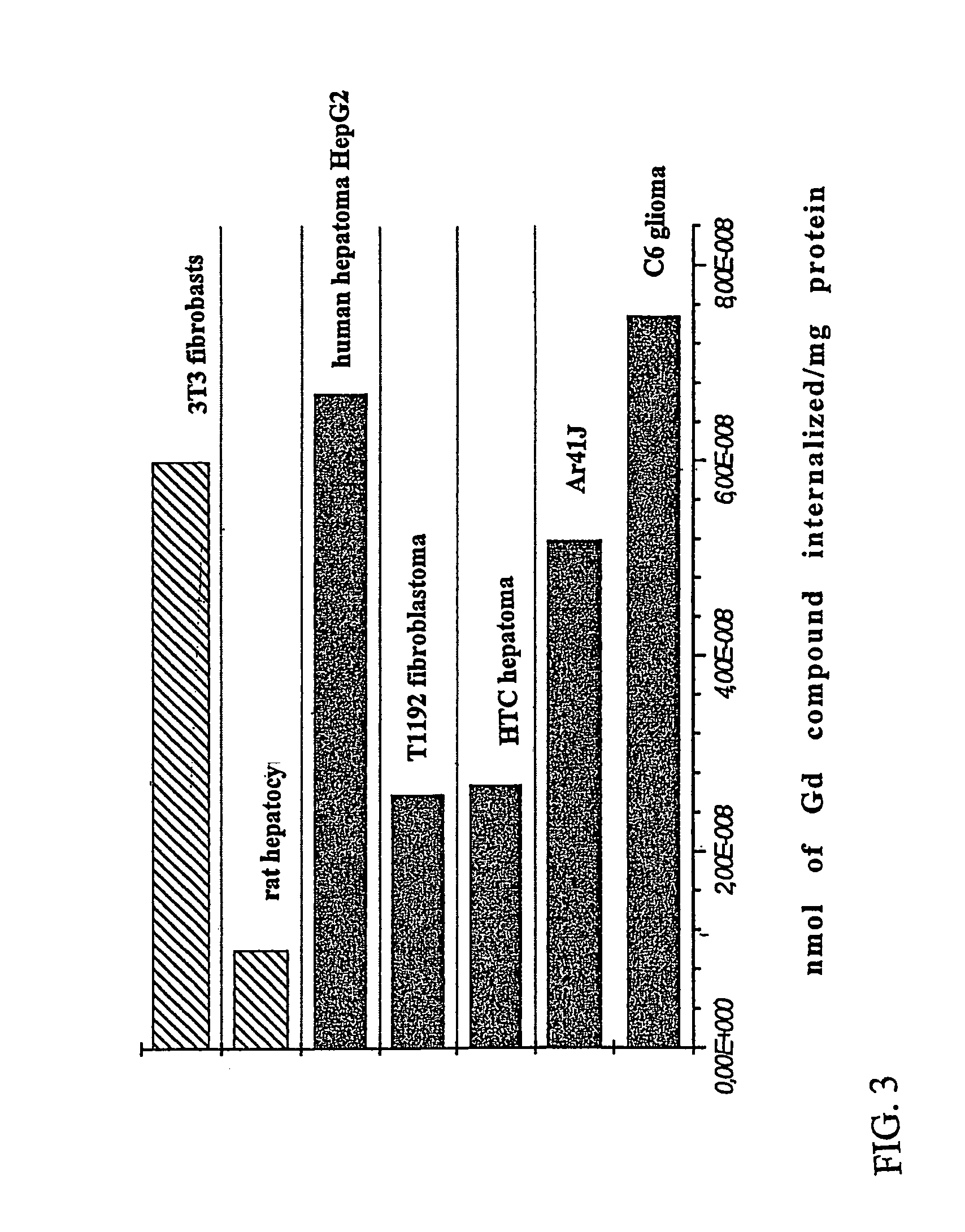
[0026]It has now been found in fact that it is possible to distinguish between tumor cells and normal cells using contrast agents based on a MRI detectable species that contain at least one MRI detectable moiety that is bound, either directly or through a spacer, to at least one molecule of a nutrient or pseudo-nutrient. The nutrient or pseudo-nutrient transporters or transporting systems that are present into the human body, will in fact recognize the nutrient or pseudo-nutrient molecule and will carry it, together with the at least one MRI detectable moiety that is linked thereto, into the cells where said MRI detectable moiety or moieties will thus concentrate to provide a more evident visualization of the cell. The altered metabolism of the tumor cells that require a much higher amount of nutrients or pseudo-nutrients or that selectively employ certain specific pseudo-nutrients, will allow a clear distinction between tumor and normal cells as the former ones will internalise a much higher amount of the compounds containing a MRI detectable moiety.

Problems solved by technology

MRI often requires the use of contrast agents, i.e. agents that influence the local relaxation behaviour of the observed nuclei in certain tissues or organs, because if MRI is performed without employing a contrast agent, differentiation of the tissue of interest from the surrounding tissues in the resulting image may be difficult.
However these metal ions are also generally highly toxic or at least poorly tolerated and need to be strongly coordinated with a ligand that occupies the major number of coordination sites.
While theoretically excellent, in practice neither of these approaches has led to a commercial product yet.
On the one hand, in fact, once the targeting MRI detectable compound is bound to the cell surface, it is not always possible to achieve or induce the desired internalization and on the other hand the use of large multimers is generally connected with an unacceptable increase in toxicity of the product because of the large molecular weight thus obtained.

Method used


Image

  • Agents for magnetic imaging method
  • Agents for magnetic imaging method
  • Agents for magnetic imaging method

Examples


example 1

Preparation of the Gadolinium Complex of 6,16-dicarbonyl-5,8,11,14,17-pentaaza-8,11,14-tricarboxymethyl-heneicosandiguadinine

(Gadolinium Complex of N,N′-(4-guanidinobutyl)diethylenetriaminopentaacetic Acid bis-amide)

[0108]

a) Preparation of Diethylenetriaminopentaacetic Acid bis-anhydride

[0109]Diethylenetriaminopentaacetic acid (10 g; 0.0255 mol) and pyridine (14.54 ml; 0.18 mol) are charged into a 100-ml reaction flask equipped with magnetic stirrer, heating oil bath, and dripping funnel. While the temperature is kept at the room value and the solution is stirred, acetic anhydride (10.56 ml; 0.11 mol) is added dropwise. The reaction mixture is heated to 65° C. for 3 hours, and then cooled to room temperature. The solid obtained is recovered by filtration on büchner, washed on filter with acetic anhydride (2×10 ml), methylene chloride (2×10 ml) and ethyl ether (2×10 ml). The white powder is then dried under vacuum yielding 8.82 g of the compound of title a) (97%).

[0110]The 1H-NMR, 13...

example 2

Preparation of the Gadolinium Complex of 6,16-dicarbonyl-5,19-dicarboxy-5,8,11,14,17-pentaaza-8,11,14-tricarboxymethyl-heneicosandioic Acid Diamide

(Gadolinium Complex of N,N′-glutamin-diethylenetriaminopentaacetic Acid bis-amide)

[0114]

a) Preparation of 6,16-dicarbonyl-5,19-dicarboxy-5,8,11,14,17-pentaaza-8,11,14-tricarboxymethyl-heneicosandioic Acid Diamide

[0115]A solution of L-glutamine (5.00 g; 0.0342 mol) in water (80 ml) is loaded into a 100-ml reaction flask and sodium hydroxide (1.37 g; 0.0342 mol) is added thereto. While keeping the reaction temperature at 15-20° C., diethylenetriaminopentaacetic acid bis-anhydride obtained as described in step a) of Example 1 (6.11 g; 0.0171 mol) is added portionwise and the reaction mixture is stirred under nitrogen atmosphere for 4 hours. The pH of the reaction mixture is then neutralised by the addition of HCl and the solvent is evaporated off under reduced pressure yielding a white product that is purified by column chromatography on Amb...

example 3

Preparation of the Gadolinium Complex of 3,6,9-triaza-3,6,9-tricarboxymethylundecanoic Acid bis-glucopyranosylamide

[0118]

(gadolinium complex of N,N′-glucosamin-diethylenetriaminopentaacetic Acid bis-amide)

a) Preparation of 3,6,9-triaza-3,6,9-tricarboxymethylundecanoic Acid bis-glucopyranosylamide

[0119]A solution of glucosamine (1.00 g; 0.00463 mol) in DMSO (10 ml) is charged into a 100-ml reaction flask and kept under stirring at 40° C. A solution of diethylenetriaminopentaacetic acid bis-anhydride obtained as described in step a) of Example 1 (0.746 g; 0.00209 mol) in DMSO (5 ml) is slowly added thereto and the reaction mixture is stirred under nitrogen atmosphere for 4 hours. Methanolic KOH is added to bring the pH of the reaction mixture to 8 and the reaction product is then precipitated by addition of methanol followed by the addition of acetone. The precipitate is washed several times with fresh acetone to remove DMSO still present and the product is then dried under vacuum. Yi...


PUM

PropertyMeasurementUnit
atomic numberaaaaaaaaaa

Abstract

The invention provides MRI detectable species of formula (I): Dp-Sn-Nm, wherein D is a MRI detectable moiety; S is a spacer; N is a molecule of a nutrient or pseudo-nutrient; n is 0 or an integer, m is an integer and p is an integer. These compounds are useful for internalizing into tumor cells an amount of the MRI detectable moiety that is distinguishably higher than the amount internalized in normal healthy cells thus allowing the diagnosis of tumors. The internalization of the MRI detectable moiety involves the nutrients or pseudo-nutrients transporting system. Preferred compounds of formula (I) are those wherein D is the chelated complex of a paramagnetic metal ion.

Description

[0001]The present invention relates to new MRI detectable species suitable for use as contrast agents in a magnetic imaging method, and to the injectable compositions containing them for the magnetic resonance imaging (MRI) of living subjects and for the diagnosing of tumors.[0002]The invention also refers to the injectable compositions of some of said new MRI detectable species for the therapy of tumors.[0003]Finally the present invention also relates to the new intermediates in the preparation of the new MRI detectable species of the invention.BACKGROUND OF THE INVENTION[0004]Magnetic resonance imaging (MRI) is a nuclear magnetic resonance technique that is used clinically to distinguish between different tissues or organs in the human or animal body through the spatial localization of water protons in the tissues or organs. The signal that is obtained by this technique and is then converted into an imaging depends in fact on the water proton concentrations and on the relaxation r...

Claims


Application Information

Patent Timeline
Patent Type & Authority Patents(United States)
IPC IPC(8): A61B5/055G01R33/28A61K41/00A61K49/00A61K49/08C07C237/10C07C237/12C07C271/22C07C279/12C07C279/14C07D257/02C07F5/00C07H5/06
CPCA61K41/009A61K41/0095A61K49/085C07C279/14C07C237/12C07C271/22C07C279/12C07C237/10
Inventor AIME, SILVIOMAINERO, VALENTINAGENINATTI, SIMONETTA CHIRICHCABELLA, CLAUDIA
Owner BRACCO IMAGINIG SPA
Why Eureka
Try Eureka