Porous biomaterials surface activation method

a biomaterial and activation method technology, applied in the field of ceramic implants and orthopedic prostheses, can solve the problems of poor quality, reduced recolonization, and associated with a certain morbidity rate, and achieve the effect of facilitating associations

Inactive Publication Date: 2012-07-10
INST NAT POLYTECHNIQUE DE TOU LOUSE +1
View PDF4 Cites 4 Cited by
  • Summary
  • Abstract
  • Description
  • Claims
  • Application Information

AI Technical Summary

Benefits of technology

[0010]The present invention aims at resolving the drawbacks of the prior art by proposing a method that makes it possible to deposit, at low temperature on biomaterials with interconnected porosity, a resorbable bioactive mineral phase, of various thickness, comprised of calcium phosphate nanocrystals analogous to bone mineral. The method according to the invention comprises an impregnation of the porous material by a suspension of bioactive calcium phosphate followed by drying under precise conditions.
[0011]This method enables the deposition of very reactive nanocrystals that considerably increase the surface reactivity of ceramics and enable the adsorption of bioactive substances capable of orienting cell activity. The main advantage of this method is to activate the surface of low specific surface and not very reactive sintered materials.
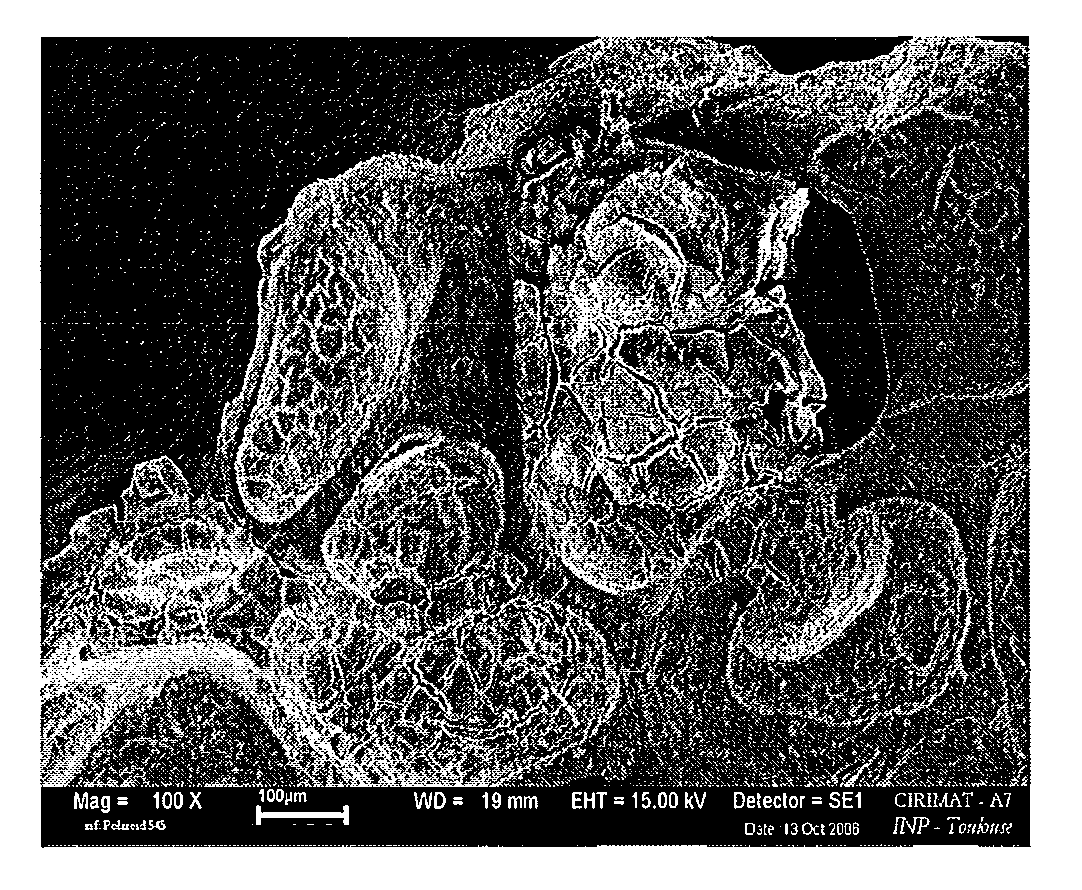
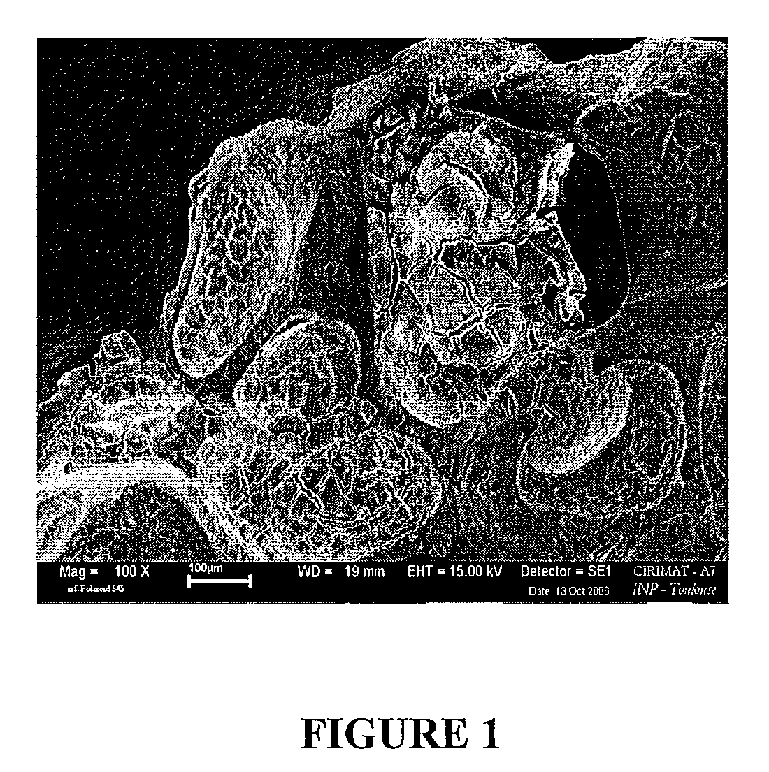
[0013]The invention thus proposes a method enabling an activation of the surface of these ceramics by a deposition of apatitic calcium phosphate analogous to bone mineral. This deposit of very high specific surface (up to 300 m2 / g) allows to increase the surface reactivity of the material and facilitates the adsorption of bioactive substances capable of orienting cell activity. This surface treatment method enables the entire surface of the ceramic to be coated, including in particular the internal surfaces of the interconnected pores of the material.
[0015]Another advantage of the method resides in the possibility of incorporating in the apatite, either at the time of its formation or after its formation, bioactive mineral ions such as strontium, magnesium, manganese, vanadate ions, etc., which can remain mobilizable and active when they are incorporated in the hydrated layer.
[0016]Similarly, the association of proteins or growth factors promoting osteogenesis or vascularization may be carried out during the coating process or instead after deposition of the nanocrystalline apatitic layer. The combination of phosphocalcic ceramics with growth factors such as Bone Morphogenetic Proteins (BMP), for example, imparts osteoinductive properties to the material [Yuan, H; Zou, P; Yang, Z; Ahang, X; De Bruijin, J D; De Groot, K; Bone morphogenetic protein and ceramic-induced osteogenesis, J. Mater. Sci. Mater. Med.; 9, 12, 717-21; 1998] and thus facilitates bone cicatrisation. The nanocrystalline apatite coating allows to improve the adsorption properties of the material vis-á-vis such proteins.
[0018]It is moreover anticipated that the enhanced properties of the nanocrystalline apatite coating with regard to the adsorption of proteins and ion exchanges may facilitate associations, directly at the surgical site, with biologically active circulating factors.

Problems solved by technology

Indeed autografts require a second surgical operation on the donor site; they only enable, in general, the filling of small volumes due to the little amount of tissue available, said tissue sometimes being of poor quality particularly in elderly patients, and they are associated with a certain morbidity rate.
Allografting leads to an often reduced re-colonization [Enneking W. F., Journal of bone and joint surgery, 73-A, 8, 1123-1141, 1991] and infection risks that can be responsible for massive osteolyses encountered in some patients.
However, its resorption rate cannot be modulated (“Bioceramics and their clinical applications”, Ed. T. Kokubo, CRC Press, 2008).
All of these ceramics are obtained by sintering at high temperature and the biological activity of such type of material is limited on account of the low specific surface of the sintered materials and their weak interaction with the various proteins and growth factors responsible for adhesion, proliferation and cell expression.
Most known nanocrystalline apatite deposition procedures make use of supersaturated calcium phosphate solutions that are difficult to use industrially, with processing times sometimes lasting several days.
These methods have moreover been rarely used to coat the inside of ceramic pores.

Method used


Image

  • Porous biomaterials surface activation method
  • Porous biomaterials surface activation method
  • Porous biomaterials surface activation method

Examples


example 1

Deposition of a Nanocrystalline Carbonated Apatite Close to Biological Apatites

[0057]Step 1: Synthesis of the carbonated apatite gel

[0058]Solution A: 48.8 g Na2HPO4.12H2O+18 g NaHCO3 in 400 ml of deionized water.

[0059]Solution B: 6.5 g CaCl2.2H2O in 150 ml of deionized water.

[0060]After complete dissolution of the salts in solutions A and B, pour solution B into solution A. Then filter and thoroughly rinse with deionized water.

[0061]Step 2: Add 50 g of gel to 200 ml of water so as to obtain a homogeneous suspension. This suspension will constitute solution C.

[0062]Step 3: A porous calcium phosphate ceramic (30 mm3 cube, 70% interconnected porosity) is immersed in solution C arranged in a vacuum flask.

[0063]A vacuum is established for around ten minutes while stirring the solution so as to eliminate any bubbles that form. Then, ambient atmospheric pressure is rapidly re-established so as to make the gel penetrate inside the pores of the ceramic. This step is repeated several times if...

example 2

Deposition of a Nanocrystalline Non-carbonated Apatite Very Rich in HPO42− Ions with a Ca / P Ratio Close to 1.35 and Having a High Proportion of Mineral Ions in the Hydrated Layer

[0070]Step 1: Synthesis of the apatite gel

[0071]Solution A: 40 g (NH4)2HPO4 in 500 ml of deionized water.

[0072]Solution B: 17.4 g Ca(NO3)2.4H2O in 250 ml of deionized water. After complete dissolution of the salts in solutions A and B, pour solution B into solution A. Then filter and thoroughly rinse with deionized water.

[0073]Step 2: Add 50 g of gel to 200 ml of water so as to obtain a homogeneous suspension. This suspension will constitute solution C.

[0074]Step 3: A porous calcium phosphate ceramic (30 mm3 cube, 70% interconnected porosity) is immersed in solution C arranged in a vacuum flask.

[0075]A vacuum is established for approximately ten minutes while stirring the solution so as to eliminate any bubbles that form. Ambient atmospheric pressure is subsequently rapidly re-established so as to make the g...

example 3

Deposition of a Nanocrystalline Carbonated Apatite with a Ca / P Ratio Close to 1.6

[0079]Step 1: Synthesis of the carbonated apatite gel

[0080]Solution A: 48.8 g Na2HPO4.12H2O+18 g NaHCO3 in 400 ml of deionized water.

[0081]Solution B: 6.5 g CaCl2.2H2O in 150 ml of deionized water.

[0082]After complete dissolution of the salts in solutions A and B, pour solution B into solution A. The suspension is left to mature for several months. Subsequently filter and thoroughly rinse with deionized water.

[0083]The other steps are identical to those of example 1.

[0084]The Ca / P ratio for these deposits (2 months' maturation) is 1.58; C / P ratio: 0.14. Crystal length: 25 nm.


PUM

PropertyMeasurementUnit
temperatureaaaaaaaaaa

Abstract

The present invention relates to a porous biomaterials surface activation method by coating with a layer of apatitic nanocrystals in order to increase their surface reactivity. The method according to the invention is characterized by the following steps: a) preparing a nanocrystalline apatitic calcium phosphate analogous to bone mineral by mixing a calcium salt solution with a phosphate salt solution in a Ca / P ratio ranging between 1.3 and 2 at a temperature ranging between 0 and 60 ° C., b) slurrying the mixture obtained in step a) in an aqueous solution so as to obtain a fluid, homogeneous paste containing 80 to 98% of water, c) bringing a porous biomaterial into contact with the suspension obtained in step b), d) drying the porous biomaterial at a temperature below 100 ° C.

Description

FIELD OF THE INVENTION[0001]The present invention relates to the field of ceramic implants and orthopedic prostheses. More specifically, it relates to a porous biomaterials surface activation method by coating with a layer of apatitic nanocrystals in order to increase their surface reactivity.BACKGROUND OF THE INVENTION[0002]Phosphocalcic ceramics first appeared in the field of biomaterials around twenty years ago. They make it possible to offset the drawbacks of biologic grafts (autografts (also known as autogenous or autologous grafts), allografts (also known as allogenic or homologous grafts) and xenografts) while promoting bone reconstructing. Indeed autografts require a second surgical operation on the donor site; they only enable, in general, the filling of small volumes due to the little amount of tissue available, said tissue sometimes being of poor quality particularly in elderly patients, and they are associated with a certain morbidity rate. Allografting leads to an often...

Claims


Application Information

Patent Timeline
Patent Type & Authority Patents(United States)
IPC IPC(8): B05D3/02A61K33/42A61F2/28A61L27/32
CPCA61L27/12A61L27/32A61L27/56C04B41/009C04B41/5048C04B41/87C04B41/4539C04B41/4549C04B35/447C04B38/00A61L2400/12A61L2400/18A61L2430/02C04B2111/00836
Inventor AUTEFAGE, HELENECAZALBOU, SOPHIECOMBES, CHRISTELEREY, CHRISTIAN
Owner INST NAT POLYTECHNIQUE DE TOU LOUSE
Why Eureka
Try Eureka