Vaccinia virus for diagnosis and therapy of tumors

a technology of vaccinia virus and tumors, applied in the field of diagnostic and pharmaceutical compositions, can solve the problems of inability to show whether, lack of sensitivity and specificity of conventional tumor diagnostic methods, etc., and achieve the effect of easy identification of such bacteria

Inactive Publication Date: 2014-02-04
GENELUX
View PDF224 Cites 27 Cited by
  • Summary
  • Abstract
  • Description
  • Claims
  • Application Information

AI Technical Summary

Benefits of technology

[0035]All of the luciferases expressed in bacteria require exogenously added substrates such as decanal or coelenterazine for light emission. In contrast, while visualization of GFP fluorescence does not require a substrate, an excitation light source is needed. More recently, the gene cluster encoding the bacterial luciferase and the proteins for providing decanal within the cell, which includes luxCDABE was isolated from Xenorhabdus luminescens (Meighen and Szittner, J. Bacteriol. 174 (1992), 5371-5381) and Photobacterium leiognathi (Lee et al., Eur. J. Biochem. 201 (1991), 161-167) and transferred into bacteria resulting in continuous light emission independent of exogenously added substrate (Fernandez-Pinas and Wolk, Gene 150 (1994), 169-174). Bacteria containing the complete lux operon sequence, when injected intraperitoneally, intramuscularly, or intravenously, allowed the visualization and localization of bacteria in live mice indicating that the luciferase light emission can penetrate the tissues and can be detected externally (Contag et al., Mol. Microbiol. 18 (1995), 593-603).
[0036]Preferably, the microorganism of the present invention is a bacterium which is not tumor-targeted, e.g. an attenuated bacterium. A list of candidate bacterial strains that might be useful for the present invention (i.e. BMPT (bacterium-mediated protein therapy) and tumor imaging) and which might not be tumor-targeted are lis

Problems solved by technology

These observations demonstrate a concomitant presence of bacteria at tumor sites; however, it was not yet clear whether the microorganisms were the cause of tumor formation or whether the tumorous tissues were more susceptible to bacterial colonization.
However, so far it could not be shown definitely whether the above discussed microorganisms are responsible for the development of disorders like tu

Method used


Image

  • Vaccinia virus for diagnosis and therapy of tumors
  • Vaccinia virus for diagnosis and therapy of tumors
  • Vaccinia virus for diagnosis and therapy of tumors

Examples


example 1

Materials and Methods

[0098](A) Bacterium Strains.

[0099]The bacterial strains used were attenuated Salmonella typhimurium (SL7207 hisG46, DEL407[aroA544::Tn10]), attenuated Vibrio cholerae (Bengal 2 Serotyp 0139, M001 DattRS1), and attenuated Listeria monocytogenes (D2 mpl, actA, plcB) The bacterial strains were kindly provided by Prof. W. Göbel (University of Würzburg, Germany).

[0100](B) Plasmid Constructs.

[0101]The plasmid pLITE201 containing the luxCDABE cassette was obtained from Dr. Marines, Biotech 24, 1998, 56-58). The plasmid pXylA-dual with the operon sequence of gfp-cDNA, lux AB, lux CD, and lux E under the control of the Xylose promoter was kindly provided by Dr. Phil Hill (University of Nottingham, UK).

[0102](C) Transformation of Bacteria

[0103]The bacteria were transformed by electroporation.

[0104](D) Tumor Cell lines. The rat C6 nitrosourea-induced glioma cell line (ATCC, Rockville, Md.) was cultured in RPMI-1640 medium (Cellgro®, Mediatech, Inc., Herndon, Va.) supplemen...

example 2

Results Obtained by Intravenous Injection of Recombinant Vaccinia Virus rVV-ruc-gfp into Mice

(A) Monitoring of Virus-Mediated Marker Gene Expression in Immunodeficient Mice

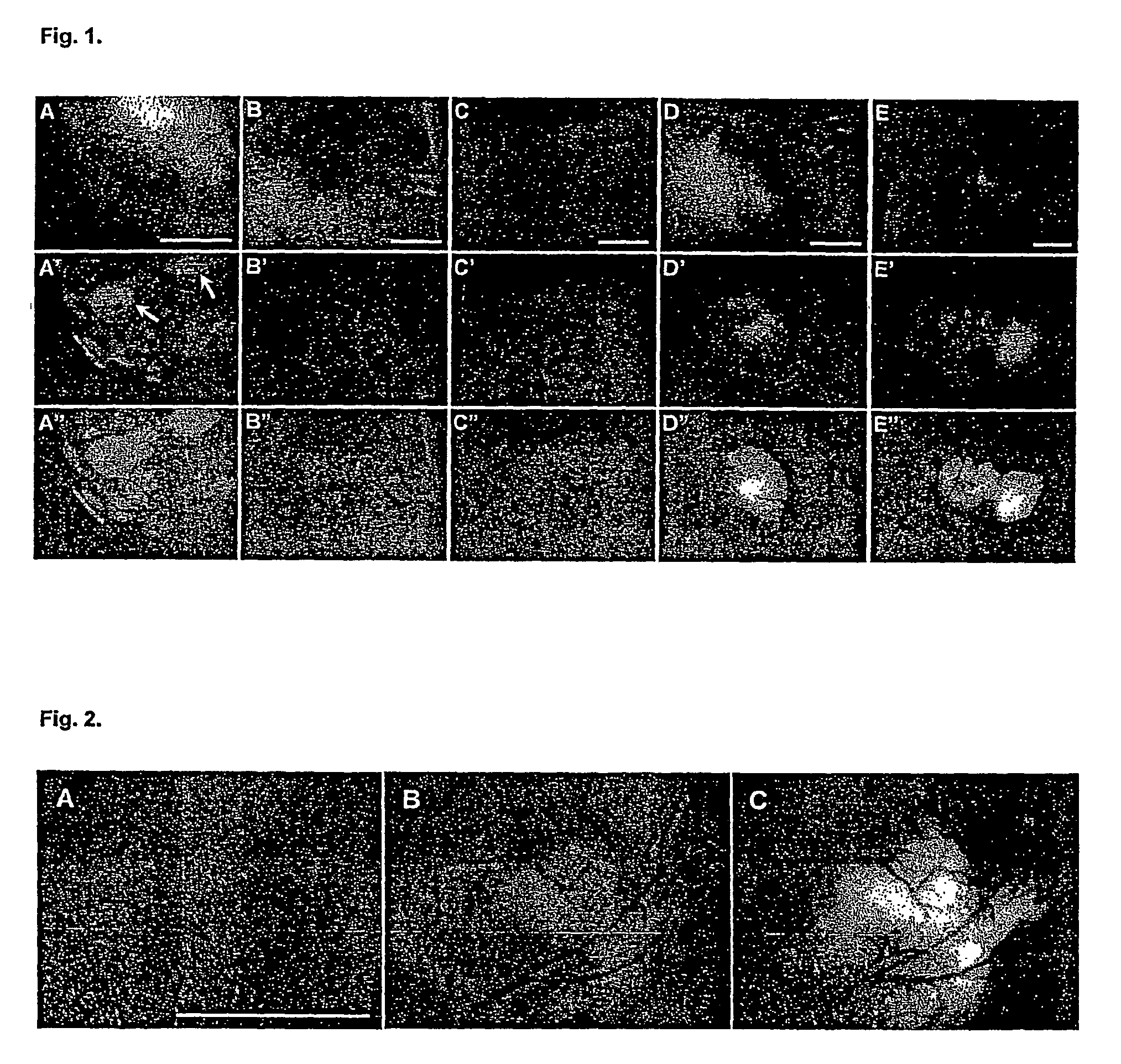
[0120]Vaccinia virus (1×10⁸ pfu) carrying the Renilla luciferase—GFP fusion expression cassette (rVV-ruc-gfp) was introduced intravenously into nude mice with no tumors. The animals were observed once every 3 days over a two-week time period under the low-light imager to monitor luciferase catalyzed light emission immediately after intravenous injection of coelenterazine, and under a fluorescence microscope to visualize GFP expression. Neither apparent luminescence nor green fluorescence was detected in the animals when imaged externally, except at certain locations that had small skin lesions. Such luminescence and fluorescence signals disappeared after a few days once the lesions had healed. Animals were sacrificed one week and two weeks after viral infection, and their organs were removed and examined for the p...

example 3

Results of Intravenous Injection of Bacterial and Mammalian Light-Emitting Cells into Mice

(A) Visualization of Light Emitting Bacteria present in Whole Animals after Intravenous Injection

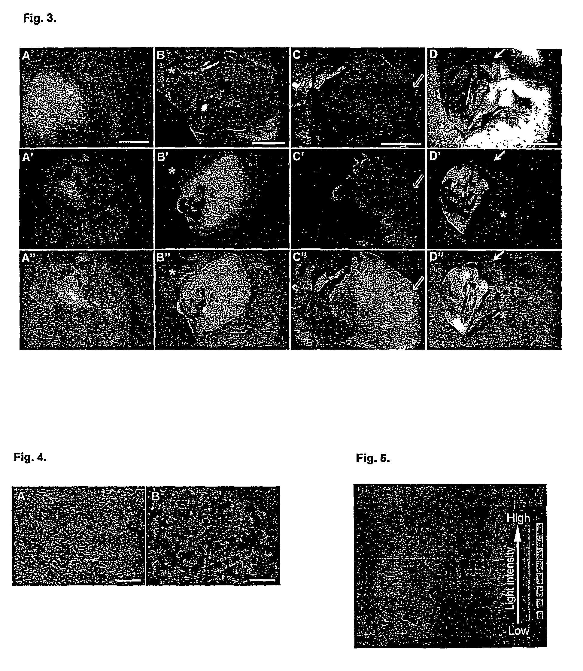
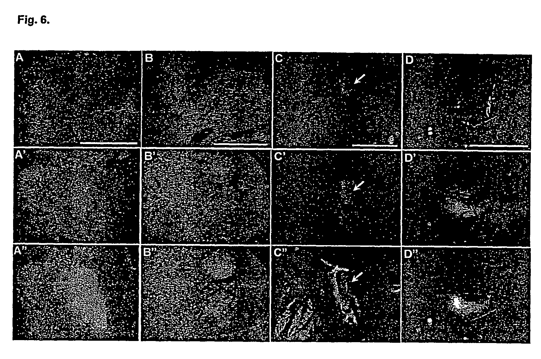
[0133]To determine the fate of intravenously injected luminescent bacteria in the animals, 107 bacteria carrying the pLITE201 plasmid in 50 μl were injected into the left femoral vein of mice under anesthesia. Following closure of the incision with sutures, the mice were monitored under the low light imager (ARGUS 100 Camera System, Hamamatsu, Hamamatsu, Japan) in real time and photons were collected for one minute. The imaging was repeated in two-day time intervals to determine the presence of light emission from a given animal. It was found that the distribution pattern of light emission following an intravenous injection of bacteria into mice was characteristic of the bacterial strains used. Injection of the attenuated V. cholera into the bloodstream resulted in light emission localized in the li...


PUM

PropertyMeasurementUnit
Temperatureaaaaaaaaaa

Abstract

Described are diagnostic and pharmaceutical compositions comprising a microorganism or cell containing a DNA sequence encoding a detectable protein or a protein capable of inducing a detectable signal, e.g. a luminescent or fluorescent protein, and, in a particular embodiment, furthermore (a) DNA sequence(s) encoding (a) protein(s) suitable for tumor therapy and/or elimination of metastatic tumors, e.g. a cytotoxic or cytostatic protein.

Description

CROSS-REFERENCE TO RELATED APPLICATIONS[0001]This application is filed under the provisions of 35 U.S.C. §371 and claims the priority of International Patent Application No. PCT / IB02 / 04767 filed Jul. 31, 2002, which in turn claims priority of European Patent Application No. 01118417.3 filed Jul. 31, 2001 and European Patent Application No. 0112591.6 filed on Oct. 30, 2001.FIELD OF THE INVENTION[0002]The present invention relates to diagnostic and pharmaceutical compositions comprising a microorganism or cell containing a DNA sequence encoding a detectable protein or a protein capable of inducing a detectable signal, e.g. a luminescent or fluorescent protein, and, in a particular embodiment, furthermore (a) DNA sequence(s) encoding (a) protein(s) suitable for tumor therapy and / or elimination of metastatic tumors, e.g. a cytotoxic or cytostatic protein.BACKGROUND OF THE INVENTION[0003]Presence of bacteria in tumors was reported approximately fifty years ago. Several publications subst...

Claims


Application Information

Patent Timeline
IPC IPC(8): A61K49/18G01N33/574A61K39/00C12Q1/66G01N33/50C12N1/21A61K48/00A61K49/00C12Q1/00A61K35/74C12Q1/70A61K35/12A61K35/76C12Q1/02C12Q1/68C12N15/03A61B5/055
CPCA61K49/0013A61K49/0097C12Q1/66A61K38/39A61K49/1896C12N2710/24132A61K38/1841C07K2319/61C07K14/78C07K14/495C07K2319/55C12Q1/02C07K2319/60A61K49/0047A61P35/00
Inventor SZALAY, ALADAR A.YU, YONG A.SHABAHANG, SHAHROKHTIMIRYASOVA, TATYANA
Owner GENELUX
Why Eureka
Try Eureka