Compositions and methods for quantitatively monitoring lipids

a quantitative monitoring and lipid technology, applied in the field of development and use of lipid sensing agents, can solve the problems of low sensitivity and robustness of methods in in situ lipid quantification, methods that do not provide quantitative information, and lipid sensors made of naturally occurring lipid binding domains may not be able to compete with endogenous cellular proteins with higher affinity

Inactive Publication Date: 2014-04-01
THE BOARD OF TRUSTEES OF THE UNIV OF ILLINOIS
View PDF0 Cites 3 Cited by
  • Summary
  • Abstract
  • Description
  • Claims
  • Application Information

AI Technical Summary

Problems solved by technology

Despite its experimental convenience and popularity, these methods do not provide quantitative information because fluorescence proteins do not undergo a spectral change upon lipid binding.
However, these methods generally suffer from low sensitivity and robustness in in situ lipid quantification.
Furthermore, lipid sensors made of naturally occurring lipid binding domains may not be able to compete with those endogenous cellular proteins with higher affinity for and / or easier access to particular lipids.
Although mass spectrometry-based lipid analysis offers higher sensitivity and provides the detailed structural information about lipids, including acyl chain compositions, the current method requires physical separation of lipids from cells and thus can provide neither spatial nor real-time temporal information.

Method used


Image

  • Compositions and methods for quantitatively monitoring lipids
  • Compositions and methods for quantitatively monitoring lipids
  • Compositions and methods for quantitatively monitoring lipids

Examples


example 1

Phosphatidylinositol-4,5-bisphosphate (PtdIns(4,5)P2) Sensor

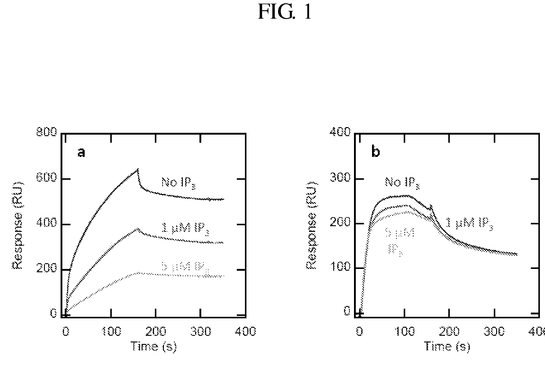
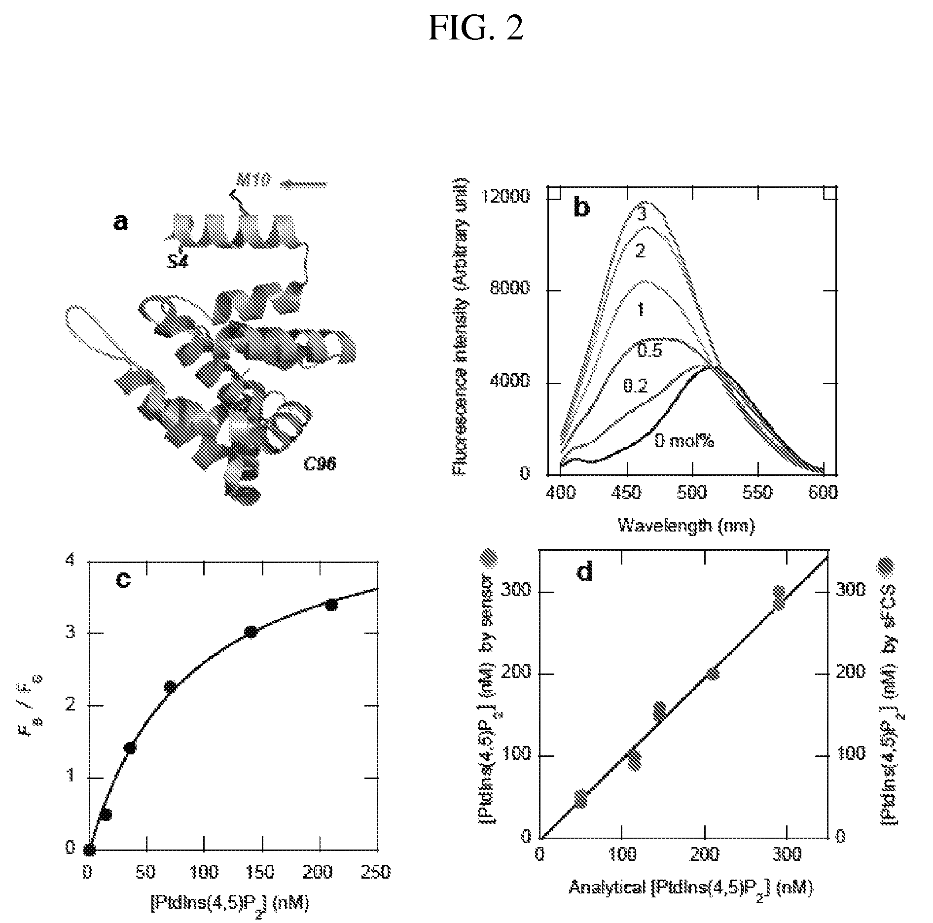
[0101]A specific sensor for PtdIns(4,5)P2, which has been implicated in numerous cell processes, including membrane remodeling and regulation of membrane proteins and cytoskeletons, was made. Although PtdIns(4,5)P2 is present mainly in the plasma membrane, its actual concentration, distribution, and spatiotemporal fluctuation has not been quantitatively determined. Traditionally, the PH domain of phospholipase Cd tagged with a fluorescent protein has been used as a cellular PtdIns(4,5)P2 probe. However, PtdIns(4,5)P2 imaging by the PH domain is known to be complicated by the ability of inositol-(1,4,5)-triphosphate, which is a hydrolysis product of PtdIns(4,5)P2, to bind the domain and displace it from the membrane. Another PtdIns(4,5)P2-selective protein was selected: the ENTH domain of epsin1 that is more stable and has lower affinity for inositol-(1,4,5)-triphosphate than the PH domain.

[0102]The ENTH domain was engineere...

example 2

Phosphatidyl Serine Sensor

[0112]The Lact-C2 gene was amplified form the EGFP-C1-Lact-C2 vector (purchased from Haematologic Technologies, Inc.) and subsequently subcloned into the pET-21a vector (Novagen). All mutations were generated by PCR mutagenesis and verified by DNA sequencing. To generate a specific turn-on sensor for PS, the DAN group was chemically incorporated to a single free cystein residue introduced to different locations of Lact-C2. Among many engineered Lact-C2 constructs, W26C-DAN showed the best optical property (See FIG. 11(a)) and was thus selected as a specific PS sensor (see FIG. 11(b)). This constructed sensor allows for in situ quantification of PS concentration in the outer (when the sensor is added to the media) and the inner (when the sensor is microinjected) faces of the plasma membrane. We were able to quantify the PS concentration on the outer plasma membrane of apoptotic Jurkat T cells that is necessary for triggering phagocytosis by macrophages. PS c...

example 3

Phosphatidic Acid Sensor

[0113]The Lact-C2 domain was converted into a PA-specific protein through protein engineering based on the crystal structure of Lact-C2 (J. Biol. Chem., (283)7230-7241 (2008)) and our molecular insight into PA and PS binding proteins. In the crystal structure, D80, H83, and Q85 interact with the serine head group of PS, so we mutated them to R, E, and K, respectively, to abrogate PS binding. Since PA has higher negative charge density without the head group, a mutant without PS binding may still interact with PA with high affinity. Our SPR analysis showed that the D80R / H83E / Q85K mutation converted the PS-specific Lact-C2 (FIG. 12(a)) into a PA-selective protein (FIG. 12(b)) and that DAN-Lact-C2-W26C / D80R / H83E / Q85K has desirable spectral properties. See FIG. 12(c). PA formation in NIH 3T3 cells in the presence of the phorbol ester, PMA, was successfully quantified.


PUM

PropertyMeasurementUnit
diameteraaaaaaaaaa
diameteraaaaaaaaaa
diameteraaaaaaaaaa

Abstract

Provided herein are fluorescent lipid binding proteins (FLBPs). The FLBPs comprise a lipid binding domain linked to a fluorophore, whrereby the fluorophore's fluorescence emission undergoes a spectral change upon lipid binding. the fluorophore is selected from the group consisting of 2-dimethylamino-6-acyl-naphthalene (DAN) and RED fluorophore and the lipid binding protein is selected from the group consisting of ENTH domain of epsin 1, C2 domain of bovine lactadherin, C 1B domain of protein kinase C-gamma, C2 domain of cytosolic phospholipase A2-beta, and PH domain of Bruton's tyrosine kinase PH.

Description

CROSS REFERENCE TO RELATED APPLICATIONS[0001]This is the national stage of International Application No. PCT / US2010 / 055606, filed on Nov. 5, 2010, which claims to the benefit of U.S. Provisional Application No. 61 / 258,959, filed on Nov. 6, 2009, the contents of which are incorporated herein by reference.FEDERALLY SPONSORED RESEARCH[0002]This invention was made with government support under grant numbers GM76581 and GM68849 awarded by the National Institutes of Health. The government has certain rights in the invention.FIELD OF THE INVENTION[0003]The present invention relates to the development and use of lipid sensing agents to quantitatively track lipids in cells.BACKGROUND[0004]Many lipids control diverse cellular processes related to cell proliferation, apoptosis, metabolism and migration. Lipids, such as phosphoinositides, sphingolipids, and fatty acids, their binding partners, and / or their downstream targets may constitute complex signaling networks that control these processes...

Claims


Application Information

Patent Timeline
Patent Type & Authority Patents(United States)
IPC IPC(8): C07K1/00
CPCG01N33/582G01N33/92G01N2405/04G01N2800/044G01N2405/06G01N33/56966
Inventor CHO, WONHWA
Owner THE BOARD OF TRUSTEES OF THE UNIV OF ILLINOIS
Why Eureka
Try Eureka