
Method for detecting and removing endotoxin

a technology of endotoxin and endotoxin, which is applied in the field of bacteriaophage tail proteins, can solve the problems of high mortality rate, endotoxin presents a problem, and inducing the death of patients

Active Publication Date: 2014-09-02
HYGLOS INVEST GMBH
View PDF34 Cites 0 Cited by
  • Summary
  • Abstract
  • Description
  • Claims
  • Application Information

AI Technical Summary

Benefits of technology

The invention aims to provide instruments and methods that can remove and detect endotoxins in solutions that have different amounts of bivalent positive ions. The technical effect of this invention is to provide a more reliable and accurate way to measure the presence of endotoxins in various solutions.

Problems solved by technology

Endotoxins effect on human and animals highly pyrogenically, so they cause fever response and are able to result in a sepsis, a heavy dysfunction of the immune system involving a high mortality rate.
Endotoxin presents a problem by genetically produced pharmaceuticals, gentherapeutic agents or substances, which are injected into humans or animals (e.g. veterinary treatment or in animal tests).
In case a drug and therein-containing proteins, respectively, have a too high endotoxin load, it is possible that this induces the death of the patient.
The misdirected immune defense damages the patient by an over-reaction.
This may induce tissue inflammation, decrease in blood pressure, tachycardia, thrombosis culminating in septic shock and multiple organ failure.
However, the removal of endotoxin on proteins, polysaccharides and DNA is problematically.
In particular, great problems exist on proteins, because of whose intrinsic properties as charge state or hydrophobicity, which almost inhibit endotoxin removal and may lead to great losses of products, respectively, during the removal process.
However particularly for proteins, there are no general applicable standard methods so far.
However, it is not qualified for high-molecular proteins or DNA.
However, the method is time-consuming because of repeating the purification process for several times.
Further, the S3 delta peptide (WO 01 / 27289) of the factor C (a component of the LAL-test) (WO 99 / 15676 both: National University of Singapore) is used for proteins (e.g. BSA, chymotrypsinogene), however this method has a low efficiency at high ionic strength and high production costs come along (production of insect cell cultures).
The recovery of this protein from the horseshoe crab or recombinant from saccharomyces is also time-consuming and cost-intensive.
Essentially three methods exist for protein solutions adapted to the properties of the target proteins in application in pharmaceutical industry:anion exchange chromatographyreserved-phase chromatography; This has the disadvantage, that it is not suitable for all proteins similarly and for hydrophobic proteins particularly problematically.
Furthermore, this method is very time-intensive and proteins are normally denaturated under the conditions of the reserved-phase chromatography, so that they have to be renaturated afterwards time-consuming and often with a high material lossRemTox (Fa.
Milipore): This method has the disadvantage that beside a very long incubation time the unspecific binding fraction is high and the recovery of proteins is often not sufficiently.
However, still the remaining concentrations of endotoxin affect toxically.
However, the precision purification is often not warranted sufficiently with the present methods.
The standard methods have relevant disadvantages and are often not applicable for certain proteins or only with a relevant loss of the target protein.
For binding reactions needing calcium, in particular buffers or additives are problematically, which coordinate calcium as EDTA, EGTA or citrate.
In addition, buffers whose salts build insoluble or hardly soluble precipitations with calcium are problematically.
However, the both first named methods are very expensive and not at least critical for nature conservation reasons because of the high demand of test animals and blood of the very rare horseshoe crab, respectively.
In fact, the LAL-test is able to be miniaturized and automated but it has massive disadvantages at the application.
It is labor-intensive, requires special trained staff, relative long incubation times, relative big sample volumes and expensive reagents.
A onetime opened LAL-solution has to be processed and used up directly, because the components aggregate within a few hours because of low stability.
Bivalent ions have to be present in the application of the test, the pH-value is relatively limited (pH 6-7.5) and present glucans often interfere the test.
The InVitro Pyrogen test requires as fresh human blood as possible and is relative time-intensive, because the production of the interleukins requires 10 to 24 hours.
The cell culture method of the company Sterogene is also, as all cell culture methods, very complex and has problems with the standardization.
Altogether, it can be fixed, that no easy manageable economic method exists for detecting endotoxin and that the currently used methods have numerous disadvantages.

Method used


Image

  • Method for detecting and removing endotoxin
  • Method for detecting and removing endotoxin
  • Method for detecting and removing endotoxin

Examples


example 1

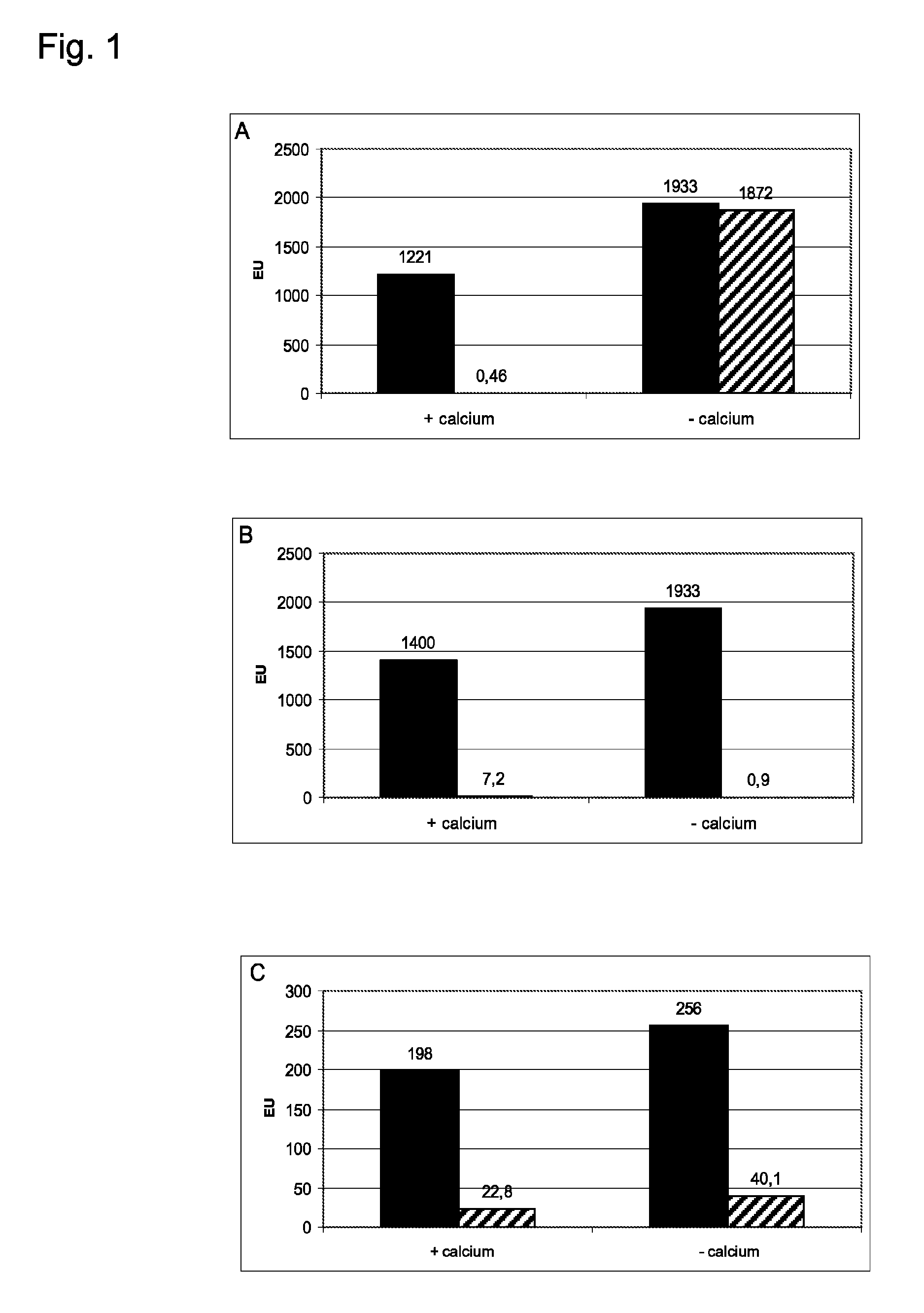
Effect of Calcium on the Endotoxin Binding of T4p12, N-Strep-Miro2p12 and N-Strep-Aeh1p12

[0143]The short tail fibre proteins T4p12, N-Strep-Miro2p12 and N-Strep-Aeh1p12 were immobilized to NHS activated sepharose 4 Fast Flow (Amersham Biosciences) according to the instruction of the manufacturer and afterwards the binding of endotoxin to said sepharoses was examined. For that purpose columns were cast, a solution with endotoxin was applied onto said columns and the flow-through was collected. The endotoxin content in the application and in the flow-through was determined via LAL-test (kinetic chromogenic LAL-test, Cambrex). The columns had volumes of 1 ml for T4p12 and N-Strep-Miro2p12 and 0.2 ml for N-Strep Aeh1p12. Each 1 ml of a BSA solution (1 mg / ml) was applied onto T4p12 and N-Strep-Miro2p12 columns and 0.2 ml of a buffer solution to N-Strep-Aeh1p12 column. The applied solutions were all studded with endotoxin of E. coli O55:B5 (approximately 1000 EU / ml). To see the effect of ...

example 2

[0145]1. Construction of Miro1, Miro2 and Effe04 with N-terminal Strep-tag: Via PCR the nucleotide sequence for the Strep-tag was added to the 5′ end of the Miro2 gene (U.S. Pat. No. 5,506,121). Therefore a primer for the 5′ end of the Miro2 gene was constructed (5′-GAA GGA ACT AGT CAT ATG GCT AGC TGG AGC CAC CCG CAG TTC GAA AAA GGC GCC GCC CAG AAT AAC TAT AAT CAC-3′; SEQ ID NO:15), which comprises the nucleotide sequence of the Strep-tag at its 5′ end (cursive in the sequence) and a restriction recognition side (NdeI, underlined in the sequence) in that way, that the gene can be inserted into the right reading frame of the expression plasmid. A primer for the 3′ end of the Miro2p12 gene (5′-CG GGA TCC TCC TTA CGG TCT ATT TGT ACA-3′; SEQ ID NO:16) was constructed, which adds a BamHI restriction recognition side behind the Miro2p12 gene (underlined in the sequence). The PCR was carried out with 35 cycles (15 s 94° C., 15 s 51° C., 1 min 74° C.). The PCR preparation was restricted wit...

example 3

[0147]Purification of N-Strep-Miro2 protein: The E. coli strain BL21(DE3) was raised with the plasmid pNS-Miro2 in a 21 agitation culture (LB-medium with ampicillin, 100 μg / ml) until a OD600 of 0.5-0.7 at 37° C. and the expression of the N-Strep-Miro2 protein was induced by the addition of 1 mM IPTG (isopropyl-β-thiogalactopyranoside). After incubation at 37° C. for 4 h the cells were harvested. Harvest cells of 10 l culture were sustained into 50 ml 10 mM sodium phosphate, pH 8.0, 2 mM MgCl2, 150 mM NaCl, disrupted by a French-Press treatment (20.000 psi) for three times and afterwards centrifuged for 30 min at 15.000 rpm (SS34). After washing for two times in the same buffer, the N-Strep-Miro2 protein was extracted from the pellet by stirring for 30 min in 10 mM TrisHCl pH 8.0, 150 mM NaCl, 1 M urea, the preparation was centrifuged for 30 min at 15.000 rpm (SS34) and the released N-Strep-Miro2 was embedded in the supernatant at 4° C. The extraction was repeated twice. The pooled s...


PUM


Abstract

The present invention relates to bacteriophage tail proteins and the derivatives and fragments thereof that are capable of binding endotoxins in the absence of bivalent positive ions, especially Ca2+ or Mg2+. Further, the present invention relates to methods for depleting endotoxins from solutions and samples using the bacteriophage tail proteins according to the present invention and to a detection method for endotoxins.

Description

CROSS-REFERENCE TO RELATED APPLICATIONS[0001]This application is a continuation of U.S. application Ser. No. 13 / 181,199, filed Jul. 12, 2011, now issued as U.S. Pat. No. 8,329,393, which is a divisional of U.S. application Ser. No. 11 / 814,358, filed Jun. 3, 2008, now U.S. Pat. No. 8,003,313, which is a national phase application under 35 U.S.C. §371 of International Application No. PCT / DE 2006 / 000098, filed Jan. 23, 2006, which claims priority to German Patent Application No.: DE 10 2005 002 969.8, filed Jan. 21, 2005. The entire text of each of the above-referenced disclosures is specifically incorporated herein by reference without disclaimer.BACKGROUND OF THE INVENTION[0002]The present invention relates to bacteriophage tail proteins and the derivatives and fragments thereof that are capable of binding endotoxins in the absence of bivalent positive ions, especially Ca2+ or Mg2+. Further, the present invention relates to methods for depleting endotoxins from solutions and samples ...

Claims


Application Information

Patent Timeline
Patent Type & Authority Patents(United States)
IPC IPC(8): C07K14/00C12Q1/00B01D15/08C07K1/13C07K1/22C07K14/005C12N7/00G01N30/00
CPCC07K14/005B01D15/08G01N30/00C07K1/13C07K1/22C12N7/00G01N2333/005C12N2795/00031G01N33/92G01N33/566G01N2400/50G01N33/56911C12N2795/00022
Inventor MILLER, STEFANMEYER, ROMANGRASSL, RENATEBIEBL, MANFREDGRALLERT, HOLGER
Owner HYGLOS INVEST GMBH
Learn More