Compositions and methods for tissue repair

a tissue and composition technology, applied in the field of tissue compositions and methods, can solve the problems of organ or tissue failure, frequent, expensive, serious problem in health care, and limited organ transplantation, and achieve the effect of facilitating the repair of the injured tissu

Active Publication Date: 2014-11-18
TRANSTARGET +1
View PDF6 Cites 24 Cited by
  • Summary
  • Abstract
  • Description
  • Claims
  • Application Information

AI Technical Summary

Benefits of technology

[0008]The present invention provides compositions and methods for the repair and / or regeneration of an injured tissue, comprising a targeted extracellular matrix derived (EMD) peptide conjugated to an injury-associated antigen-specific binding component for the repair and / or regeneration of an injured tissue. The targeted EMD peptide can provide for a surface at the injured tissue whereby cells can attach and grow, thereby facilitating the repair of the injured tissue in vivo.
[0009]The present inventors have surprisingly discovered that EMD peptides ranging in size from about 8 amino acids to about 100 amino acids can mimic the properties of full length proteins from which the EMD peptides are derived, as described herein. These short peptides are advantageous because they are easy to manufacture, have reduced immunogenicity, lower toxicity, show greater homogeneity and reproducibility in conjugation to an injury-associated antigen-specific binding component, and are easier and more cost effective to manufacture as pharmaceutical compositions as compared to the full-length proteins or longer peptides as disclosed herein.

Problems solved by technology

Organ or tissue failure remains a frequent, costly, and serious problem in health care despite advances in medical technology.
These treatments, however, are not perfect solutions.
Organ transplantation is limited by the availability of donors and complications such as tissue rejection.
Surgical reconstruction is costly, highly invasive, and not always effective.
Mechanical devices cannot functionally replace an organ, for example, dialysis machines can only help to remove some of the metabolic waste from the body.
Likewise, maintaining drug concentration levels in vivo, comparable to the control systems of the body, are difficult to achieve.
Finally, tissue engineering while promising, is encumbered by size limitations that typically require the assembly of the engineered tissue in vitro, followed by surgical implantation in vivo.

Method used


Image

  • Compositions and methods for tissue repair
  • Compositions and methods for tissue repair
  • Compositions and methods for tissue repair

Examples


example 1

Cell Attachment Assay

Reagents

[0119]Immulon 1B, untreated 96-well plates (CoStar), and untreated 35 mm dishes (CoStar) were purchased from Fisher Scientific (Pittsburg, Pa.). Human umbilical vein endothelial cells (HUVECs), cell culture media and supplements were purchased from Lonza (Basel, Switzerland). EMD peptides as shown in SEQ ID Nos: 1-4, were synthesized by Commonwealth Biotechnologies Inc., (Richmond, Va.). Amino acid analysis was performed on the peptides to verify the amino acid sequence.

Cell Attachment Assay

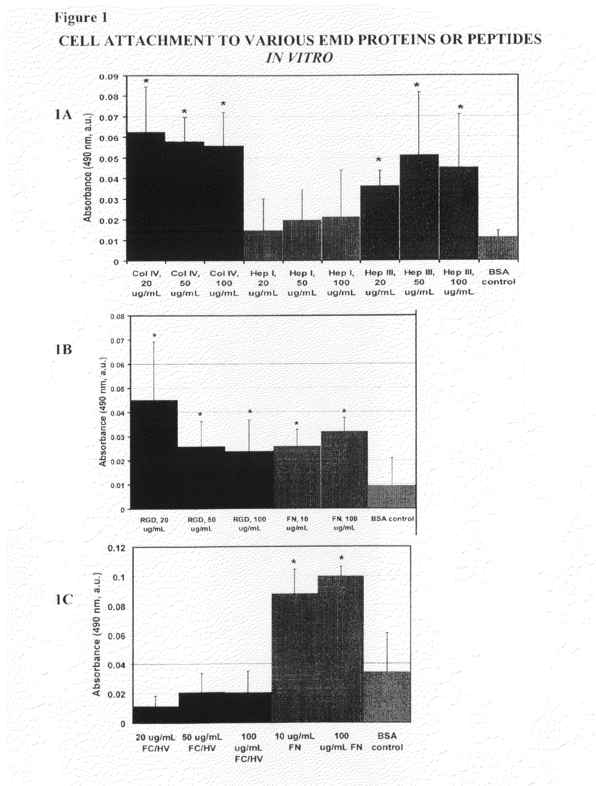
[0120]For the adhesion studies 96-well Immulon 1B plates were used. Various concentrations of the EMD peptides (SEQ ID Nos: 1-4) were tested for their ability to bind cells, and compared to the full-length protein, from which the EMD peptides were derived as positive controls. Bovine Serum Albumin (BSA) treated wells were used as a negative control. Full-length collagen IV, and the EMD peptides Hep I, Hep III, FCHV, and RGD were tested at concentrations of 20 μg / ml, 5...

example 2

Cell Proliferation Assay

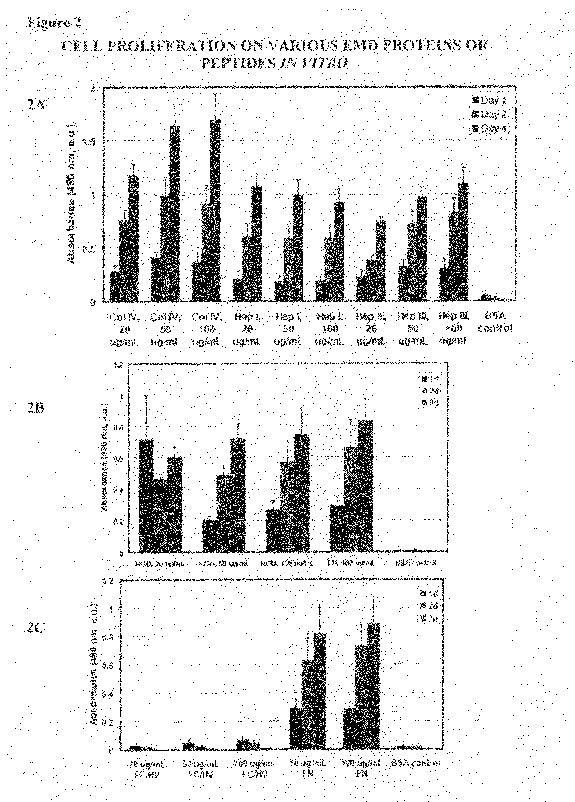
[0122]To determine if EMD peptides can activate cells by stimulating cellular proliferation, 96-well plates coated using the same protocol as described above for the cell attachment assay was used. Following treatment of the wells, HUVEC cells (Lonza, Basel Switzerland) were then added to the wells at a concentration of 5×10³ to 1×10⁴ cells per well and incubated under standard culture conditions for 1, 2, 3 or 4 days. Cell proliferation was then determined at the end of the incubation period using an MTS tetrazolium / formazan assay (Promega, Madison, Wis.) according to the manufacturer's instructions.

[0123]As shown in FIG. 2, full length proteins Col IV and fibronectin and EMD peptides Hep I, (SEQ ID NO:1) Hep III (SEQ ID NO:2), and RGD (SEQ ID NO:4) all showed increased cellular proliferation compared to cells cultured in wells coated with BSA. Notably, cells cultured in wells coated with FCHV (SEQ ID NO:3) was not different from that of cells cultured in we...

example 3

Cell Migration Assay

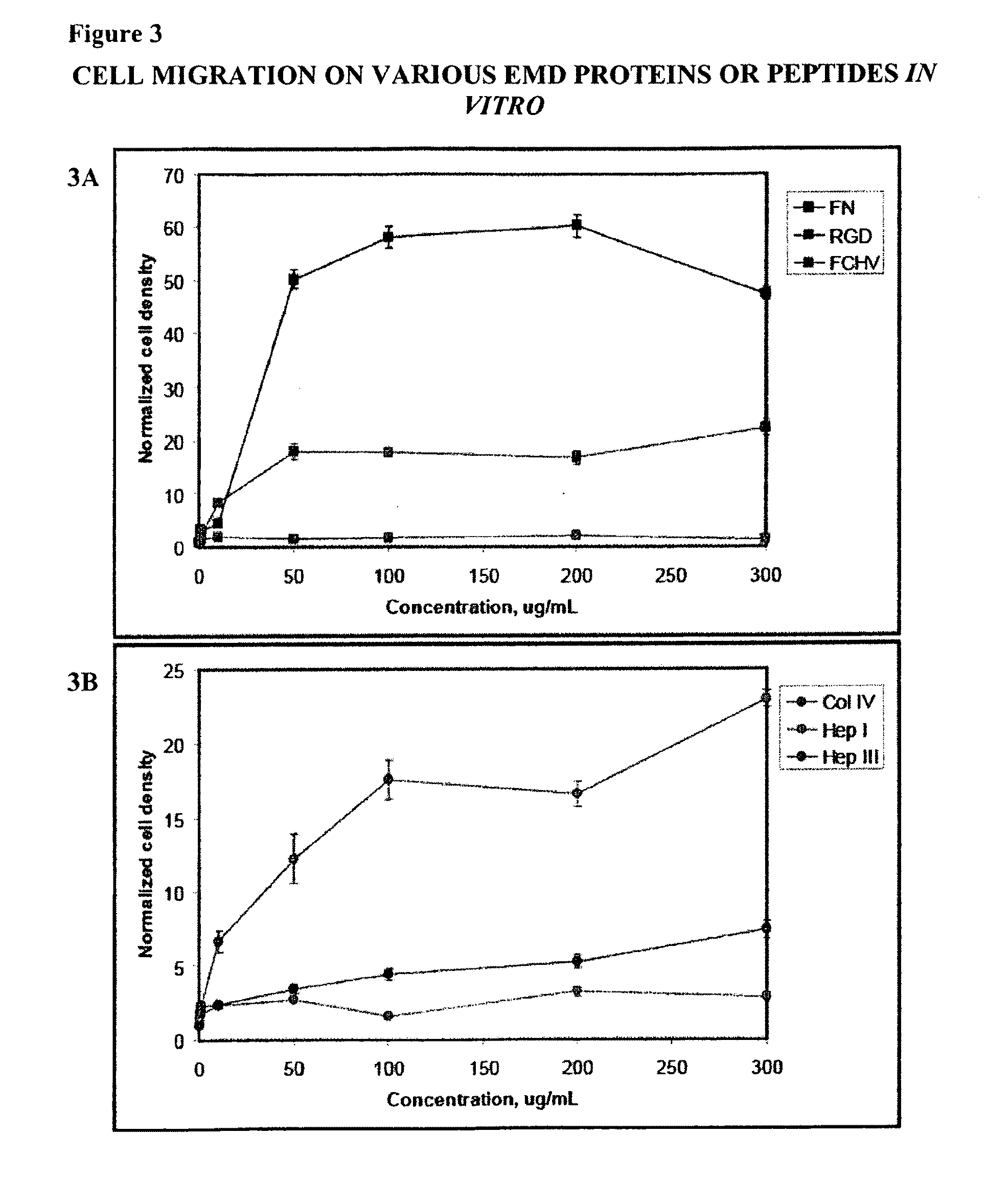
[0124]In order to test the ability of EMD peptides to recruit cells haptotactic migration assays with Hep I (SEQ ID NO:1), Hep ITT (SEQ TD NO:2), FC / HV (SEQ ID NO:3), RGD (SEQ ID NO:4), FN, and Col IV were conducted using a modified Boyden chamber. Haptotactic migration was performed in triplicate and was assessed using a modified Boyden chamber (Corning, CoStar, Acton, Mass.). The assay was carried out as follows: the lower chamber was first blocked with 10% BSA for at least 30 minutes at 37° C. followed by 3 washings with PBS. The lower surface of the membrane on the upper chamber was coated with approximately 10 μL of protein or EMD peptide solution having a concentration between 500 ng / mL-300 μg / mL and allowed to incubate for 15-30 minutes at 37° C. and then allowed to air dry at room temperature under aseptic conditions. At least one assay was performed for each concentration. Basal cell media supplemented with 0.5% BSA was then added to the lower chamber an...


PUM

PropertyMeasurementUnit
concentrationsaaaaaaaaaa
pHaaaaaaaaaa
body weightaaaaaaaaaa

Abstract

The present invention provides compositions and methods for targeting an extracellular matrix derived (EMD) peptide predominantly to an injured tissue, as opposed to an uninjured tissue in vivo. The targeted EMD peptide facilitates the repair and / or regeneration of the injured tissue by providing a surface for cells to attach and grow, thereby facilitating the repair and / or regeneration of the injured tissue.

Description

CROSS-REFERENCES TO RELATED APPLICATIONS[0001]This application claims benefit of U.S. Provisional Patent Application No. 60 / 941,051, filed on May 31, 2007, the disclosure of which is hereby incorporated by reference.FIELD OF THE INVENTION[0002]This invention relates to compositions and methods for the repair and / or regeneration of an injured tissue by targeting an extracellular matrix derived (EMD) peptide to the injured tissue.BACKGROUND OF THE INVENTION[0003]Organ or tissue failure remains a frequent, costly, and serious problem in health care despite advances in medical technology. Available treatments include organ transplantation, surgical reconstruction, mechanical devices (e.g. pace makers and kidney dialysis machines), drug therapy, and tissue engineering. These treatments, however, are not perfect solutions. Organ transplantation is limited by the availability of donors and complications such as tissue rejection. Surgical reconstruction is costly, highly invasive, and not a...

Claims


Application Information

Patent Timeline
Patent Type & Authority Patents(United States)
IPC IPC(8): C07K14/52C07K16/00A61K39/44A61K47/48C07K14/47C07K16/28C12N9/64C07K14/78C07K14/715A61K39/00C07K14/705
CPCA61K47/48415C07K14/70553C07K2319/00C07K16/2833C07K14/70542C07K14/4716C07K14/70564C07K14/4721C12N9/6475C07K14/52A61K47/48538C07K14/70525C07K14/7151C07K14/78A61K38/00C07K7/08A61K2039/505A61K47/6811A61K47/6843A61K47/6849A61P9/10C07K2318/00A61K47/68
Inventor LEE, RANDALL J.MIHARDJA, SHIRLEYHUANG, MANLEYLARRICK, JAMES W.
Owner TRANSTARGET
Why Eureka
Try Eureka