Method for expansion of human corneal endothelial cells
a corneal endothelial cell and human corneal technology, applied in the field of corneal endothelial cell expansion, can solve the problems of insufficient supply of donor corneas, gradual decrease of the cell population with age, and irreversible corneal edema
- Summary
- Abstract
- Description
- Claims
- Application Information
AI Technical Summary
Benefits of technology
Problems solved by technology
Method used
Image
Examples
example 1
Preparation of Human Corneal Endothelial Stem Cells from Limbal Biopsy for Culture
[0050]A sector of peripheral endothelial layer was obtained by biopsy from the same eye or the other eye of the patient being treated or from another living individual during a trabeculectomy procedure. The eye lid was sterilized with povidone-iodine. Under sterile conditions, a conjunctival flap was created for about 8 mm in length with a limbal base about 5 mm to about 6 mm from the limbus. A limbal base of a scleral flap having a dimension of about 5 mm in length and about 400 μm in depth was created about 3 mm from the limbus, and the flap was extended to the cornea and about 2 mm from the limbus. An anterior chamber was entering from the limbal area and extending for about 3 mm in length parallel around the limbal zone when the sclerocorneal flap was lifted. Trabeculectomy was performed and corneal endothelial layer was separated from the removed tissue. The removed corneal endothelium was then us...
example 2
Preparation of Human Corneal Endothelial Stem Cells from Donor Cornea
[0051]A sector of peripheral corneal endothelial layer was also obtained from a donor cornea. A sclerocorneal rim of the donor cornea was obtained when the central cornea was trephined for penetrating keratoplasty. The corneal endothelial layer with Descemet's membrane was separated carefully from the trabecular meshwork with surgical blade No. 15. Six sectors of peripheral corneal endothelial layer were obtained from one donor cornea.
[0052]The sector of peripheral corneal endothelial layer containing corneal endothelial stem cells was placed in a 35 mm dish containing 1.5 ml of culture medium. The culture medium was OPTIMEM-1 (purchased from Invitrogen, San Diego, Calif., USA) as a basal medium, supplemented with 5% to 8% patient's serum, and 10 ng / ml of Fibroblast Growth Factor (FGF), 5 ng / ml Epidermal Growth Factor (EGF), 10 μg / ml of ascorbic acid, 50 μg / ml penicillin, 50 μg / ml streptomycin, 2.5 mg / ml fungizone....
example 3
Preparation of Human Amniotic Membrane and Expansion of Human Corneal Endothelial Stems Cells on the Human Amniotic Membrane
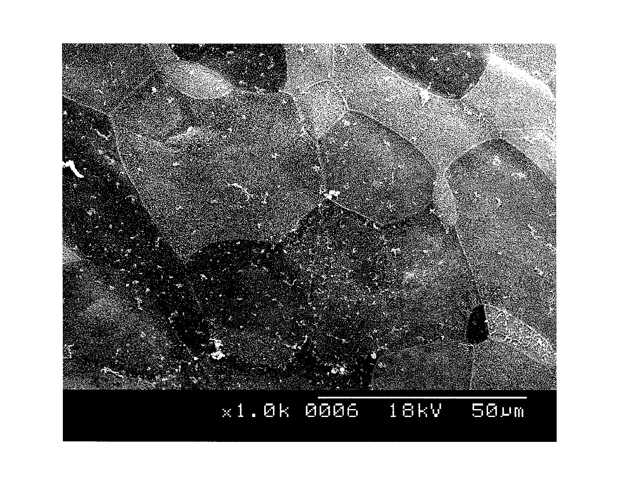
[0053]In accordance with the tenets of the Declaration of Helsinki and with proper informed consent, a human amniotic membrane was obtained at the time of cesarean section. The human amniotic membrane was washed with PBS containing antibiotics (5 ml of 0.3% ofloxacin) and then stored in DMEM and glycerol at −80° C.
[0054]The amniotic membrane, with basement membrane side up, was affixed smoothly onto a culture plate and placed at 37° C. under 5% CO2 and 95% air, in a humidified incubator overnight before use. The corneal endothelial explant culture was performed on the amniotic membrane. The peripheral corneal endothelial layer containing corneal endothelial stem cells was planted or transferred onto the basement membrane side of the amniotic membrane in a 35 mm dish containing about 1 ml to about 1.5 ml of the culture medium described above. The medium was chan...
PUM
| Property | Measurement | Unit |
|---|---|---|
| thickness | aaaaa | aaaaa |
| diameter | aaaaa | aaaaa |
| size | aaaaa | aaaaa |
Abstract
Description
Claims
Application Information
 - R&D
- Intellectual Property
- Life Sciences
- Materials
- Tech Scout
- Unparalleled Data Quality
- Higher Quality Content
- 60% Fewer Hallucinations
Browse by: Latest US Patents, China's latest patents, Technical Efficacy Thesaurus, Application Domain, Technology Topic, Popular Technical Reports.
© 2025 PatSnap. All rights reserved.Legal|Privacy policy|Modern Slavery Act Transparency Statement|Sitemap|About US| Contact US: help@patsnap.com

