

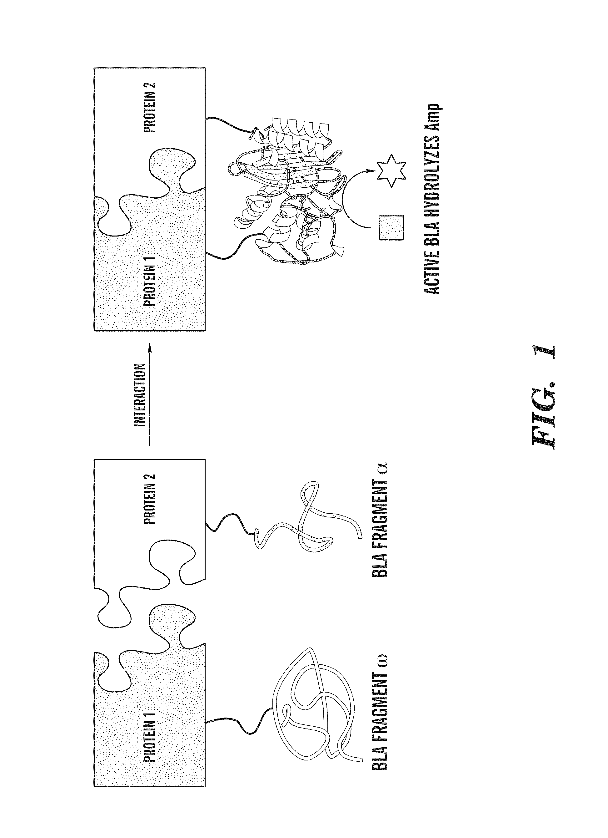
System useful for reporting protein-protein interactions in the bacterial periplasm
a protein-protein interaction and periplasm technology, applied in the field of system useful for reporting protein-protein interactions in the bacterial periplasm, can solve the problems of complicated interpretation of interaction data, high false positive rate of both y2h and b2h gal4-type assays, and inability to select genes
- Summary
- Abstract
- Description
- Claims
- Application Information
AI Technical Summary
Benefits of technology
Problems solved by technology
Method used
Image

Examples
example 1
Construction of Vectors
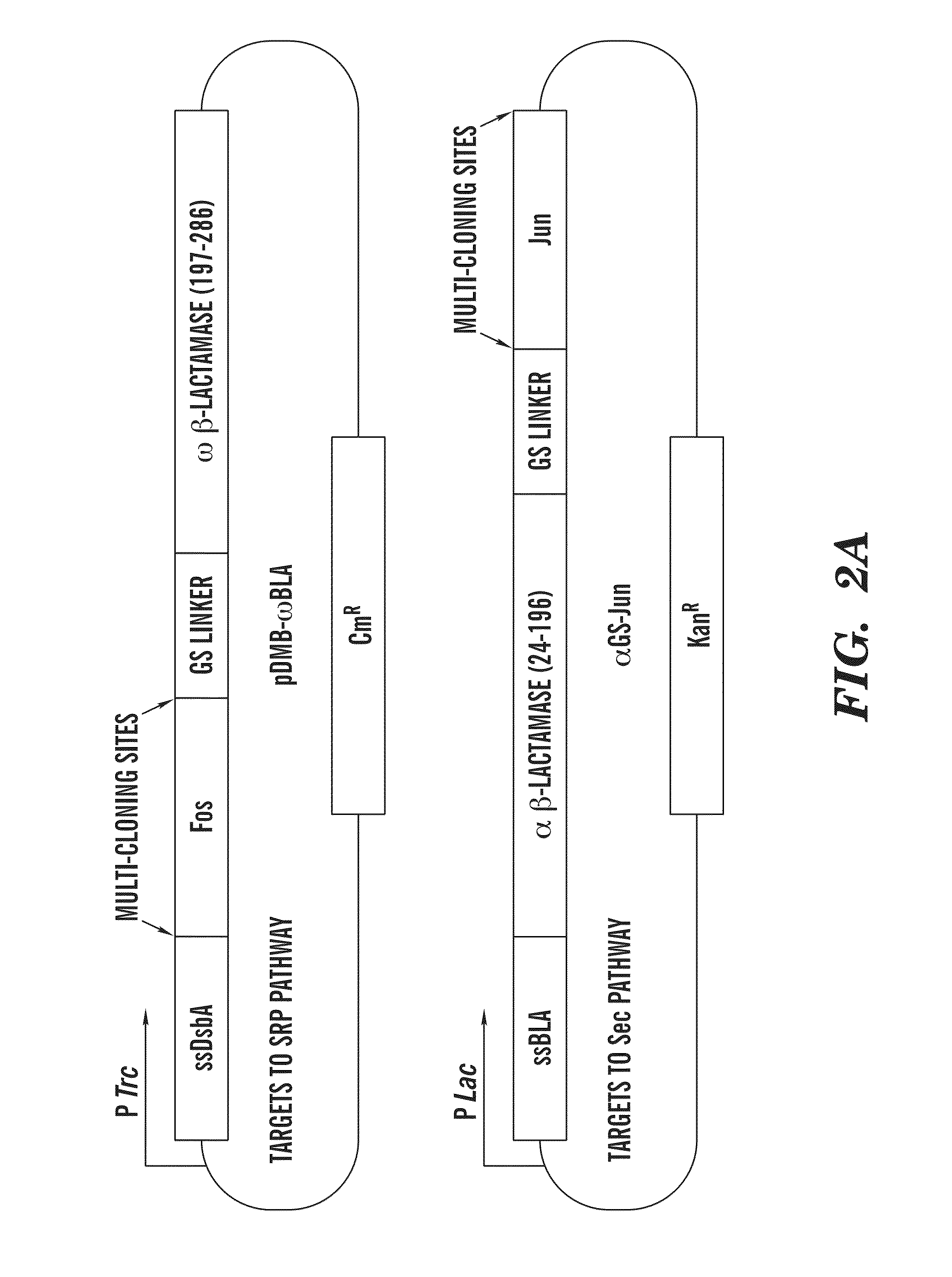
[0075]All cloning was performed using standard molecular biological techniques. A gene sequence comprising a (GGGGS)3-NGR linker sequence followed by residues 198-286 of TEM-1 BLA was cloned between the BamHI and HindIII sites of pDMB to create pDMB-ωBLA. Genes to be fused to the ωBLA fragment were then cloned into this vector between XbaI and SalI sites. Genes to be fused to the αBLA fragment were cloned between the KpnI and BamHI sites of vector αGS-Jun (Wehrman, T., et al., “Protein-protein Interactions Monitored in Mammalian Cells Via Complementation of Beta-lactamase Enzyme Fragments,”Proc Natl Acad Sci USA 99:3469-3474 (2002), which is hereby incorporated by reference in its entirety). Fos and Jun leucine knockouts and scFv-GCN4 were as in (DeLisa, M. P., “Versatile Selection Technology for Intracellular Protein-protein Interactions Mediated by Bacterial Hitchhiker Transport Mechanism,”Proc Natl Acad Sci USA 106:3692-3697 (2009), which is hereby incorpor...
example 2
Expression of Fusion Proteins and Cell Growth Assays
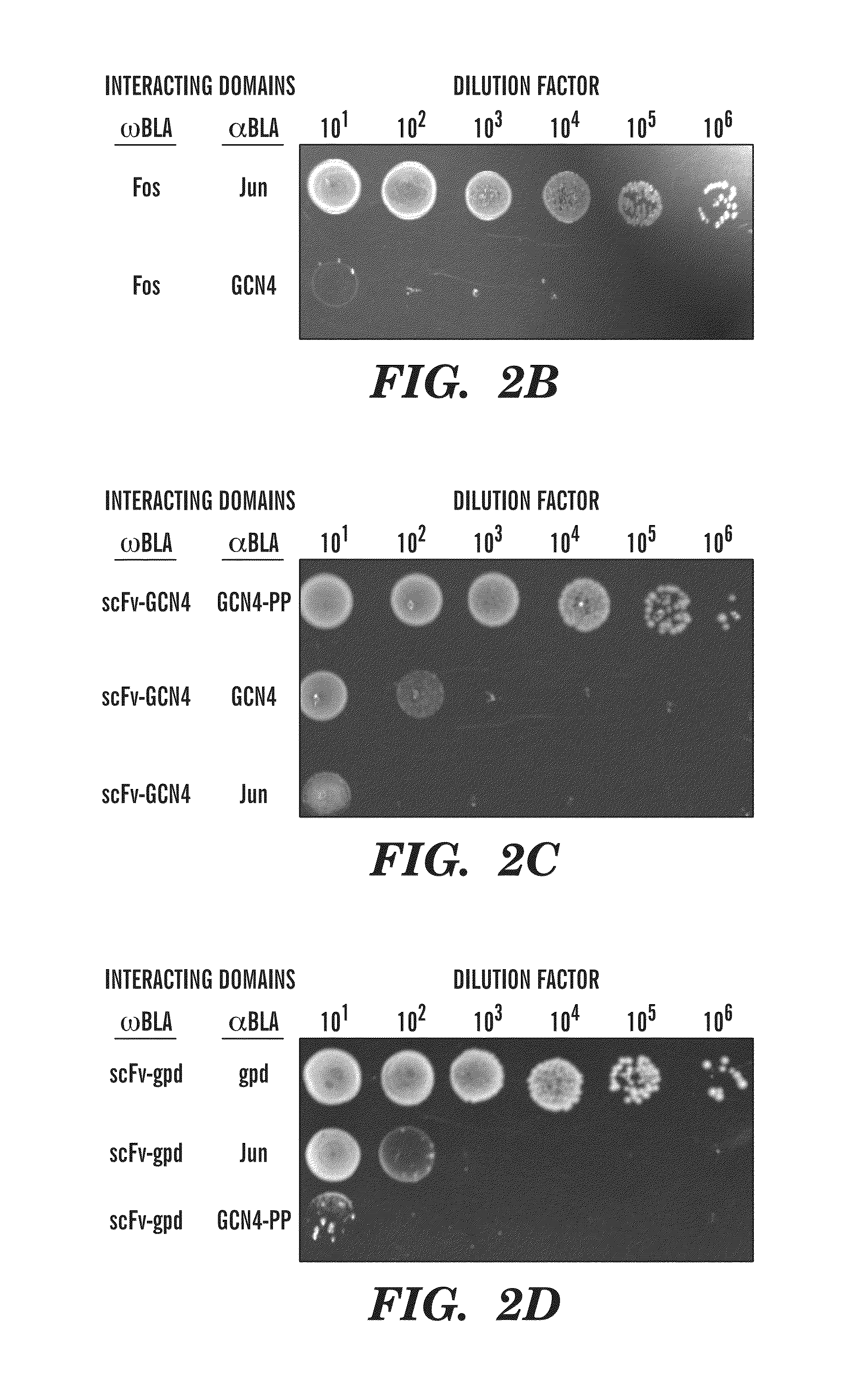
[0076]E. coli MC4100 cells were co-transformed with plasmids pDMB-P1-ωBLA (CmR) and aGS-P2 (KanR), where P1 and P2 represent proteins of interest. Cells were grown overnight at 37° C. in LB medium containing 20 μg / mL chloramphenicol (Cm) and 50 μg / mL kanamycin (Kan). Screening of cells on LB agar was performed by first normalizing overnight cultures by OD600 and then spotting 5 μL, of serially diluted (10-106-fold) cells on LB agar plates with 12, 25, 50, or 50 μg / mL of ampicillin (Amp). In all cases, the plates were incubated 16 h at 37° C. and then imaged using a ChemiDoc System (BioRad). For MBC / MIC determination, approximately 200 colony forming units (CFUs) of each clone were plated on LB agar plates containing 0, 3, 6, 12, 25, 50, 100, 200, 400, 800, or 1600 μg / mL Amp or 20 μg / mL Cm. The minimum bacteriocidal concentration was determined as the minimum Amp concentration at which no colonies appeared on the plates. Minimum inh...
example 3
Antibody-Antigen Interactions are Reported in Two Cases
[0078]To determine the ability of the assay of the present invention to detect and eventually engineer antibody-antigen interactions, interacting protein pairs as well as non-interacting proteins were cloned into two plasmids carrying the two inactive Bla fragments (FIG. 2A). In this study, two antibody-antigen pairs were examined. First, an scFv which is known to bind the GCN4 leucine zipper was tested. In addition to the scFv and wild-type GCN4 pair, a double proline mutant, GCN4-PP as an antigen for the scFv was also tested. GCN4 is normally a homodimer, but a double proline mutant of GCN4 (GCN4-pp) exhibits reduced dimerization, thereby reducing the amount of inactive α / α dimers in the assay. FIGS. 2B-D show the results of spot plating of the interacting pairs, with α-Jun as a negative control. The spot plates correspond to a 16-fold higher MBC (minimum bacteriocidal concentration, see Example 2 for full definition of MIC / MB...
PUM
| Property | Measurement | Unit |
|---|---|---|
| concentration | aaaaa | aaaaa |
| concentration | aaaaa | aaaaa |
| concentration | aaaaa | aaaaa |
Abstract
Description
Claims
Application Information