Apparatus and method for implantation of devices into soft tissue

a technology of soft tissue and implantation method, which is applied in the field of apparatus and methods for the surgical implantation into soft tissue of devices, can solve the problems of high device failure rate, over-design of devices with larger-than-needed foreign materials, and functional failures, and achieve the effect of reducing the damage to soft tissu

Active Publication Date: 2016-08-09
UNIVERSITY OF PITTSBURGH
View PDF22 Cites 3 Cited by
  • Summary
  • Abstract
  • Description
  • Claims
  • Application Information

AI Technical Summary

Benefits of technology

[0028]The present invention describes an apparatus and method for implanting devices into soft tissue with accuracy and precision in three dimensions as well as in prescribed insertion speed and trajectory, and reduces the damage that occurs to the soft tissue into which the device is implanted.
[0029]The invention apparatus comprises several sub-systems that provide the functionality to achieve accuracy, precision and damage reduction. These subsystems are: 1. an actuator, such as the M272 piezo motor sold by PI of Auburn, Mass., that moves at a controlled high velocity along a single, longitudinal axis (i.e the implantation trajectory) with a large travel range up to 50 mm and better than 20 micron positional accuracy; 2. an actuator, such as that made from K-740 PZT by Piezo Technologies, Indianapolis, Ind. that can impart an oscillatory motion at frequencies between 18 kHz and 30 kHz in two directions, corresponding to the transverse directions to the insertion direction, or the single, longitudinal axis) for reduction of insertion forces; 3. a load cell, such as the Sensotec Model 31 sold by Honeywell of Morristown, N.J., that measures the force between the device being implanted and the tissue surface during implantation; 4. a contact sensor for accurate detection of the point and time of contact between the soft tissue and the device being implanted, which can be achieved by monitoring the electrical characteristics of the piezo-actuator described in subsystem 2; 5. a laser ranging system, like the Hokuyo URG-04LX-UG01 with a Sokuiki sensor, for referencing the position and motion of the tissue with respect to the insertion system and the device being implanted; 6. an imaging system, such as the SE-1008-400X video microscope from Selectech Electronics of Guangdong, China, for identifying the optimal insertion location to minimize mechanical damage to tissue vasculature; 7. a clamping mechanism, such as an MGP800 series clamp from Sommer Automatic of Ettlingen, Germany, to hold the device being implanted, that is operated in coordination with the actuator; 8. a set of clamping surfaces with a design that is customized to the form of the device being implanted; 9. a cartridge for holding multiple devices with a design that is customized to the form of the device being implanted; 10. a dispenser that moves devices from the cartridge to the clamp, with a design that is customized to the form of the device being implanted, or an operating sequence in which the cartridge is stationary and the actuator and clamp execute a predefined sequence to move to the next device to be implanted and pick it up in the clamp; and 11. the action of the system and its various subsystems are coordinated using software such as Labview from National Instruments of Austin, Tex. which can provide a graphical user interface for ease of use, data acquisition from the various subsystems for real-time monitoring of the insertion procedure and offline analysis for diagnostic and clinical evaluation. A software like Matlab's Image Processing module from Mathworks of Natick, Mass., can provide the capability of capturing and manipulating image data and processing them according to a variety of algorithms that identify sensitive tissue structures, overlay images of the implantation sites and compute overlap area of sensitive tissue structures and implantation sites.

Problems solved by technology

Furthermore, a critical issue is to ensure that implantation occurs satisfactorily; that is, the device is inserted in at the required depth without device failure.
Manual insertions of devices cannot provide this level of control in positioning and insertion, therefore leading to high rate of device failure during insertion, over-design of devices with larger-than-needed foreign materials, and functional failures.
Two key issues are 1) the inability of current interfaces to reliably obtain accurate information from tissue over a period of decades, and 2) currently measured signals from tissue cannot be reliably used to control high degree-of-freedom (DOF) prostheses with high speed and resolution.
Failure of biological soft tissue interfaces may be caused by several issues.
Tissue reaction with the probe results in encapsulation that insulates the electrode by impeding diffusion of chemical and ionic species and may impede current flow from the soft tissue to the interfaces.
Lastly, interconnects may fatigue and break due to stresses.
Experiments in animals have resulted in some neural electrode sites failing while others keep working for several years.
However, reliable and manufacturable connections to the braided wires become difficult when scaled to arrays.
The low velocity of manual insertions, either by hand or using stereotaxic frames, results in observable soft tissue dimpling prior to penetration of the tissue.
Dimpling was found to be accompanied by soft tissue compression that resulted in damage to the tissue and reduced signal extraction.
An adverse effect of the mechanism is recoil of the endpiece due to the return spring which can lead to retraction of the interface device if it remains adhered to the endpiece.
This technique does not allow for accurate placement of the interface in soft tissue because there is no visibility of the contact points between the interface and the tissue.
They found the impact between the endpiece and the backside of the interface often led to damage of the interface, so they added a “footplate” to the device.
However, because the device is not mechanically connected to a fixed reference structure, it is subject to elastic recoil from the soft tissue into which it is implanted and this can lead to retraction of the interface from the tissue.
The literature reports other insertion mechanisms of varying levels of complexity and functionality.

Method used


Image

  • Apparatus and method for implantation of devices into soft tissue
  • Apparatus and method for implantation of devices into soft tissue
  • Apparatus and method for implantation of devices into soft tissue

Examples


Embodiment Construction

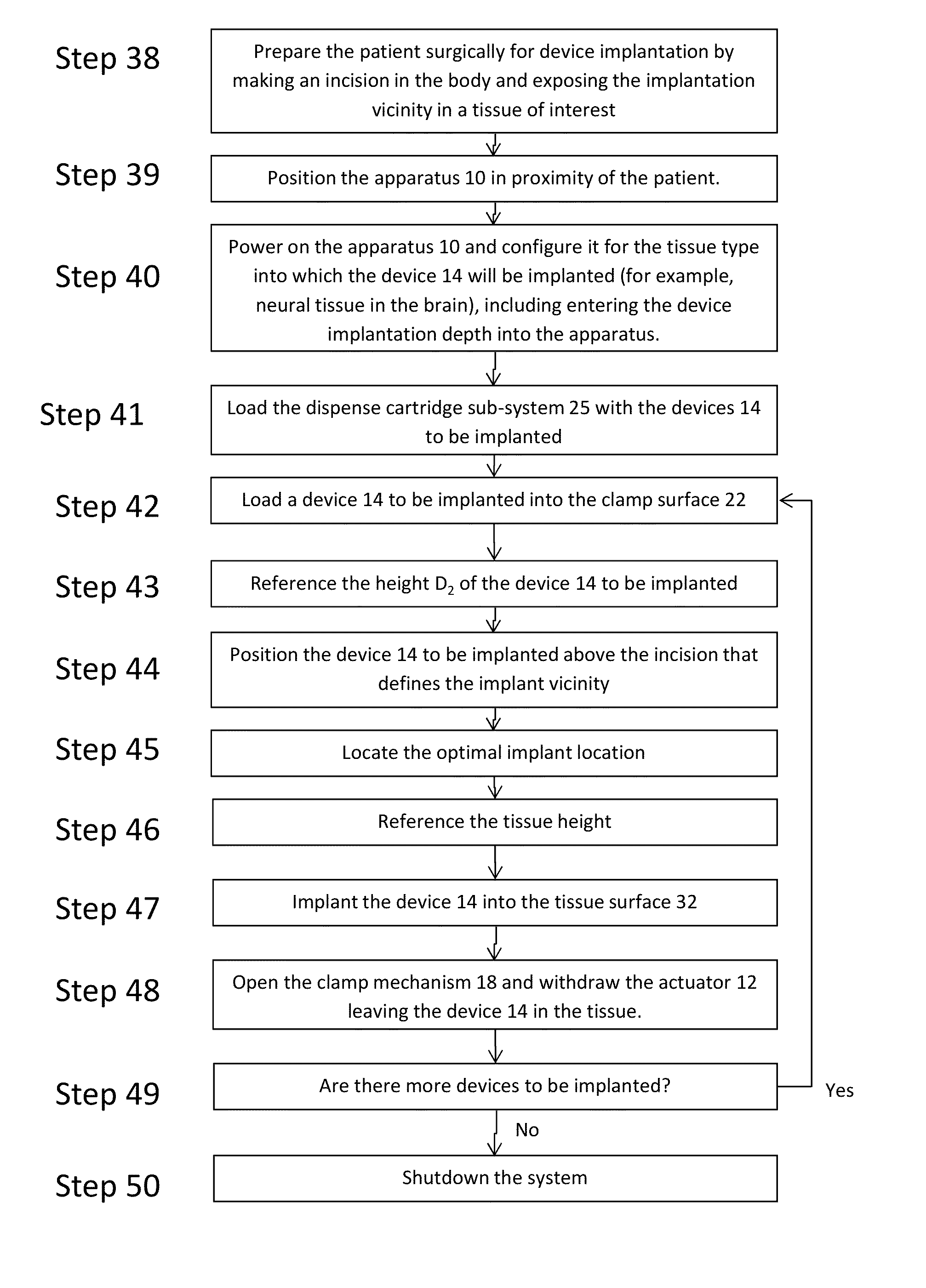
[0045]The present invention addresses the problem of the accurate and precise placement and depth of devices implanted into human tissue with reduced soft tissue damage. Now turning to FIGS. 5 and 14, the present invention is an apparatus 10 for holding, referencing, targeting, and implanting devices of various sizes, shapes and materials into soft tissue and the method that must be followed for the achievement of accuracy, precision and reduced damage using the apparatus. The apparatus 10, by way of representation and not invention limitation, can include an actuator 12, laser ranging sub-system 30, contact sensor 16, load cell 20, imaging sub-system 28, clamp mechanism 18 with clamp surface 22, processor 40, memory 42, and display 44 for implantation of device 14 into tissue surface 32 (FIG. 6B) of a patient. The high level operation of the apparatus and the method for executing implantations is detailed in the process flow diagram in FIG. 13. The step numbers are labels and are n...


PUM


Abstract

Apparatus and method for surgeon-assisted rapid surgical implantation of devices into soft tissue. The apparatus comprises several subsystems that enable the referencing of the spatial position and orientation of the device being implanted with respect to the soft tissue into which it is being implanted and then the controlled implantation of the device at a predefined speed with higher positional accuracy and precision and a reduction in soft tissue damage, provided by ultrasonic assisted motion, compared to current state-of-the-art implantation methods and devices. The method includes automated loading of the device being implanted into a clamping mechanism from a cartridge holding a number of implants, referencing of the device position and orientation, referencing of the surface of the tissue into which the device is being implanted, monitoring of the tissue motion, identification of desirable implant location based on the soft tissue profile, allowance of surgeon selection and fine adjustment of the final implant location, high-speed implantation, device release and implant actuator retraction.

Description

CROSS-REFERENCE TO RELATED APPLICATIONS[0001]This application is a Non-provisional application that claims the benefit of U.S. Provisional Application Ser. No. 61 / 690,044, titled APPARATUS AND METHOD FOR IMPLANTATION OF DEVICES INTO SOFT TISSUE, filed Jun. 18, 2012, which is incorporated by reference herein.STATEMENT REGARDING FEDERALLY-SPONSORED RESEARCH AND DEVELOPMENT[0002]This invention was made with partial government support under DARPA grant N660011114025. The government has certain rights in this invention.FIELD OF THE INVENTION[0003]The invention relates to apparatus and methods for the surgical implantation into soft tissue of devices, such as (1) prosthetic neural interfaces between computers and the machinery they control and biological tissue, for example neurons and the nodes of Ranvier on axons in nerve bundles, (2) optical fibers for the localized stimulation of neurons and other cell types, and (3) drug delivery catheters, among others. The micrometer-scale interfac...

Claims


Application Information

Patent Timeline
Patent Type & Authority Patents(United States)
IPC IPC(8): A61B5/00
CPCA61B5/6838A61B5/6843A61B5/4851A61B2090/064A61B34/30A61B34/20A61B34/70A61B90/10
Inventor GILGUNN, PETER J.OZDOGANLAR, O. BURAKKOZAI, TAKASHI DANIEL YOSHIDAFEDDER, GARYCUI, XINYANWEBER, DOUGLAS J.
Owner UNIVERSITY OF PITTSBURGH
Why Eureka
Try Eureka