Screening method for therapeutic agent for chondropathy and modified chondrocyte for treatment of chondropathy

a technology of chondropathy and chondrocyte, which is applied in the direction of drug compositions, skeletal/connective tissue cells, instruments, etc., can solve the problems of hypertrophy of cartilage-like cells induced from stem cells, and achieve the effect of reducing cartilage volume and reducing cartilage volum

Active Publication Date: 2017-02-07
KYOTO UNIV
View PDF5 Cites 0 Cited by
  • Summary
  • Abstract
  • Description
  • Claims
  • Application Information

AI Technical Summary

Benefits of technology

[0015]The screening method of the present invention allows screening for a substance useful as a therapeutic agent for a disease associated with reduced cartilage volume and / or a disease associated with impaired cartilage growth. The modified chondrocyte of the present invention is useful for the amelioration of a disease associated with reduced cartilage volume and / or a disease associated with impaired cartilage growth. The modified chondrocyte of the present invention can also be used, as a disease model cell that reproduces the disorder of chondrocyte differentiation, in the analysis of the clinical conditions of chondropathy and in the screening for a drug for chondropathy.

Problems solved by technology

Due to little repair capacity of cartilage, there is no radical cure for osteoarthritis and current therapy is aimed to control pain by limiting exercise and using an analgesic.
However, BMPs have the effect of, after increasing cartilage, hypertrophying the chondrocytes and replacing them with bone, which causes problems in use for the treatment of articular cartilage.
However, cartilage-like cells induced from stem cells tend to hypertrophy, which poses problems (Non Patent Literature 5).

Method used


Image

  • Screening method for therapeutic agent for chondropathy and modified chondrocyte for treatment of chondropathy
  • Screening method for therapeutic agent for chondropathy and modified chondrocyte for treatment of chondropathy
  • Screening method for therapeutic agent for chondropathy and modified chondrocyte for treatment of chondropathy

Examples


reference example 1

Analysis of Chondrocyte Differentiation in SIK3 Knockout Mice

(1) SIK3 Knockout Mice (Hereinafter Referred to as “SIK3 KO Mice”)

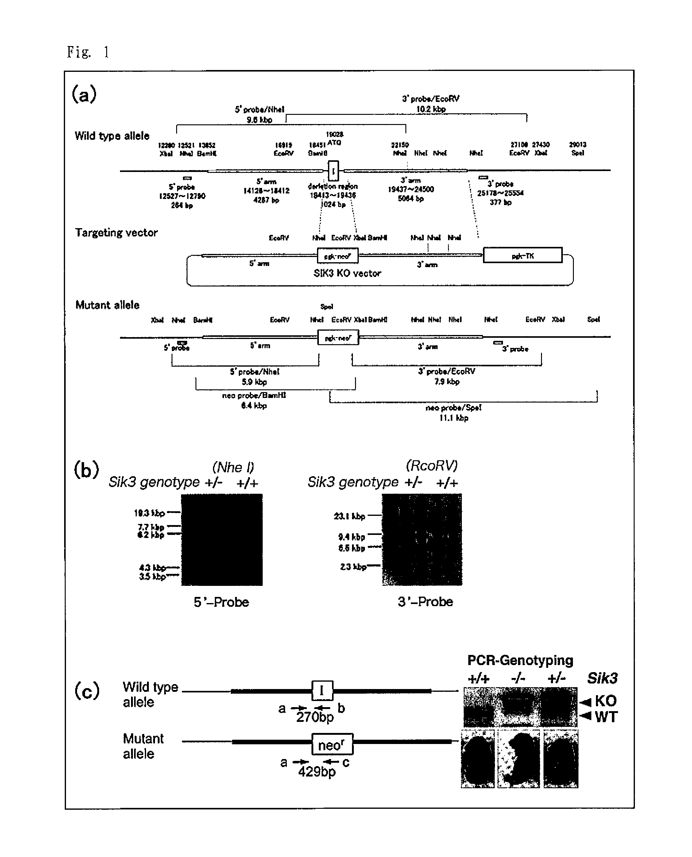
[0068]A targeting vector carrying, in consecutive order from the 5′ end to 3′ end, the 5′-end genome sequence of the mouse SIK3 gene (5′ arm), a pgk promoter, a neomycin resistance gene, and the 3′-end genome sequence of the mouse SIK3 gene (3′ arm) (see FIG. 1 (a)) was generated in accordance with a usual method. The obtained targeting vector was introduced into mouse ES cells and the cells were cultured in G418-containing medium to generate neomycin-resistant ES cells. DNA was extracted from the obtained neomycin-resistant ES cells and homologous recombinant clones were identified by southern blot analysis (see FIG. 1 (b)).

[0069]The obtained homologous recombinant clones were microinjected into blastocysts obtained from BDF2 mice, and the manipulated blastocysts were implanted into the womb of mice. The mice that conceived after the implantation gave birth...

reference example 2

Rescue Test Using SIK3 Transgenic Mice

(1) SIK3 Transgenic Mice (Hereinafter Referred to as “SIK3 Tg Mice”)

[0078]A transgene construct was generated by linking a human SIK3 cDNA (Accession No. NM_025164) to a promoter / enhancer for the α2(XI) collagen chain gene (Col11a2), which directs cartilage-specific expression (Tsumaki N, et al. J Cell Biol. 1996; 134: 1573-1582). This transgene insertion was cut from the vector backbone, purified, and then microinjected into mouse fertilized eggs. The eggs were implanted into the oviduct of pseudo-pregnant mice. From the mice produced from the pseudo-pregnant mice, SIK3 Tg mice were selected and thus a SIK3 Tg mouse line was established. The genotype of the fetuses or the newborns was confirmed by PCR using the genomic DNA extracted from the tail or ear tissue. The following primers were used for the PCR.

[0079]

(SEQ ID NO: 7)Forward primer: 5′-GATGTCGGATGCAGTTCTC-3′(SEQ ID NO: 8)Reverse primer: 5′-ATGTCTGTAATACACGTAGATGGATA-3′

(2) Test Method

[008...

reference example 3

Observation of Growth Plate of Knee Joint of SIK3 Transgenic Mice

[0084]Three-, six-, and eight-month-old SIK3 Tg mice and wild-type (WT) mice were euthanized by carbon dioxide inhalation and dissected for harvesting the forelimbs and hindlimbs. The harvested limbs were fixed in formalin and then decalcified in EDTA. Next, sections were produced and stained in the same manner as in Reference Example 1.

[0085]The obtained specimens were observed under an optical microscope. FIG. 6 shows histologically stained sections including the knee joint of each of the 3-, 6-, and 8-month-old mice. The left is the wild-type mice (“Wt” in the figure) and the right is the SIK3 Tg mice (“LI-hSIK3 Tg” in the figure). As is apparent from FIG. 6, the growth plate was observed in both of the wild-type mice and the SIK3 Tg mice at 3 months old, but the growth plate disappeared from the SIK3 Tg mice at 6 and 8 months old, while the growth plate in the wild-type mice was observed at 6 and 8 months old.

[0086...


PUM

PropertyMeasurementUnit
volumeaaaaaaaaaa
stiffnessaaaaaaaaaa
Tgaaaaaaaaaa

Abstract

The use of salt-inducible kinase 3 allows screening for a substance having an effect of increasing cartilage volume and / or an effect of normalizing chondrocyte differentiation. A modified chondrocyte in which the expression or function of salt-inducible kinase 3 gene is inhibited or enhanced can be used to ameliorate a disease associated with reduced cartilage volume and / or a disease associated with impaired cartilage growth.

Description

TECHNICAL FIELD[0001]The present invention relates to a screening method for a therapeutic agent for chondropathy and a modified chondrocyte for the treatment of chondropathy.BACKGROUND ART[0002]Osteoarthritis (OA) is a progressive degenerative joint disease characterized by the degeneration of articular cartilage and the growth and reconstitution of subchondral bone. The symptoms are stiffness, restriction of the movement, pain, and the like and the incidence is higher with increasing age. Due to little repair capacity of cartilage, there is no radical cure for osteoarthritis and current therapy is aimed to control pain by limiting exercise and using an analgesic. If the symptoms have progressed and the disease has reached the end stage, artificial joint replacement is performed to excise degenerative articular cartilage and cover the site with a metal cover.[0003]In an attempt to develop a cartilage repair promoter, compounds or proteins for promoting the growth of chondrocytes ha...

Claims


Application Information

Patent Timeline
Patent Type & Authority Patents(United States)
IPC IPC(8): G01N33/48G01N33/50G01N33/487G01N33/483C12Q1/48C12N5/077C12Q1/68G01N33/68G01N33/53A61K35/00A61K35/32A61P19/08A61P43/00C12N15/09C12Q1/6897G01N33/15
CPCC12Q1/6897C12N5/0655C12Q1/485C12N2501/727G01N2500/02G01N2500/10A61P19/08A61P19/10A61P43/00
Inventor TSUMAKI, NORIYUKITAKEMORI, HIROSHI
Owner KYOTO UNIV
Why Eureka
Try Eureka