Methods of generating corneal cells and cell populations comprising same

a corneal cell and cell technology, applied in the field of generating corneal cells, can solve the problems of letting light pass through relatively uninhibited, limiting the method, and living donors at risk of limbal stem cell deficiency

Inactive Publication Date: 2017-02-21
INST NAT DE LA SANTE & DE LA RECHERCHE MEDICALE (INSERM) +1
View PDF3 Cites 1 Cited by
  • Summary
  • Abstract
  • Description
  • Claims
  • Application Information

AI Technical Summary

Problems solved by technology

However, the collagen fibers are arranged in a parallel pattern, allowing light waves to constructively interfere, thus letting light pass through relatively uninhibited.
This method is further limited due to a shortage of donors.
Further, living donors are at risk of developing limbal stem cell deficiency.
This technology does not allow treating patient immediately following injury, since it requires the cultivation of the cells for several weeks prior to grafting.
Moreover, allogenic limbal stem cell transplantations to a patient are eventually rejected.

Method used


Image

  • Methods of generating corneal cells and cell populations comprising same
  • Methods of generating corneal cells and cell populations comprising same
  • Methods of generating corneal cells and cell populations comprising same

Examples


example 1

Differentiation of Human Embryonic Stem Cells to Corneal Cells

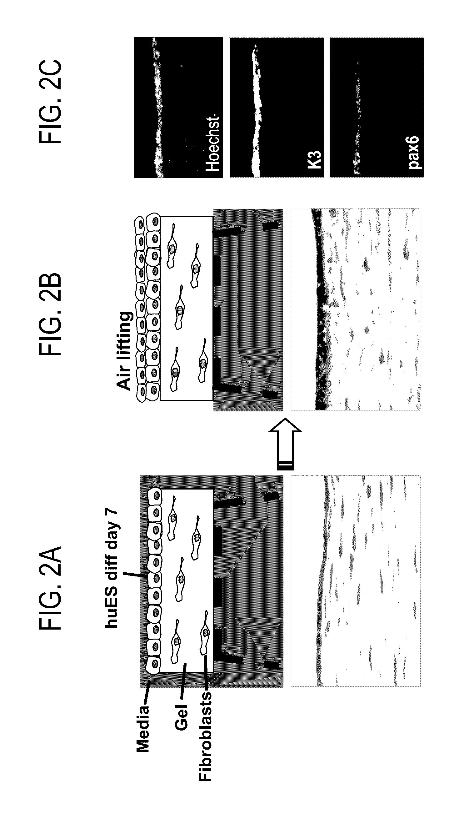
[0162]I. Preparation of corneal fibroblasts conditioned epithelial medium: To prepare conditioned media, primary corneal fibroblasts were extracted from human cornea that is not suitable for transplantation by incubation of the cornea with Dispase II (2 mg / ml) overnight at 37° C. The epithelial sheet was removed and the fibroblasts were cultured in culture dishes in dulbeco's modified eagle medium (DMEM) supplemented with 10% new born calf serum. When the fibroblast cell population reached 80%-100% density (see FIG. 2A), cell proliferation was stopped by incubating in mitomycin C (8 μg / ml) for three hours. To obtain conditioned media, the mitomycinized cells were incubated with epithelial media ((DMEM 60%, HamF12 30%, FCII 10%, insulin 5 μg / ml, hydrocortisone 0.5 μg / ml, EGF 10 ng / ml, 0.2 mM Adenine, 10 nM Cholera toxin). The medium was collected every day and replaced by fresh media for up to 10 days. The medium was filte...

example 2

Generation of Corneal Tissue from Embryonic Stem Cells

[0173]Materials and Methods

[0174]After 1-2 weeks of differentiation of pluripotent stem cells on collagen IV or matrigel in the presence of conditioned media, cells were collected by dispase or trypsin and reseeded onto a corneal stroma equivalent gels in a 3D organotypic reconstitution assay: primary human corneal fibroblast cells were embedded in gels that were composed of matrigel and collagen I, and hES-derived corneal cells were seeded on top of these gels in epithelial media. Corneal stratification was induced by air-liquid interphase as illustrated in FIGS. 2A-C and further exemplified in Gaggioli et al., Nature cell biology volume 9, number 12, 2007. Then air liquid interface was induced by reducing media inside the inserts and cells were allowed to stratify for 1-2 weeks, while medium was replaced every day. Alternatively, differentiated cells were seeded on human amniotic membrane and lifted from the culture (as describ...

example 3

Differentiation of Human Induced Pluripotent Stem (iPS) Cells to Corneal Cells

[0175]I. Preparation of corneal fibroblasts conditioned epithelial medium: as described in Example 1.

[0176]II. Coating of culture dishes: as described in Example 1.

[0177]III. Differentiation of iPS cells into corneal epithelial lineage: iPS derived from human hair follicle cells or dermal fibroblasts were seeded on collagen IV or matrigel-coated dishes. Medium was replaced every second day by fresh corneal fibroblast conditioned epithelial media for about two weeks. On days 3, 8 and 14, BMP-4 was added to the medium.

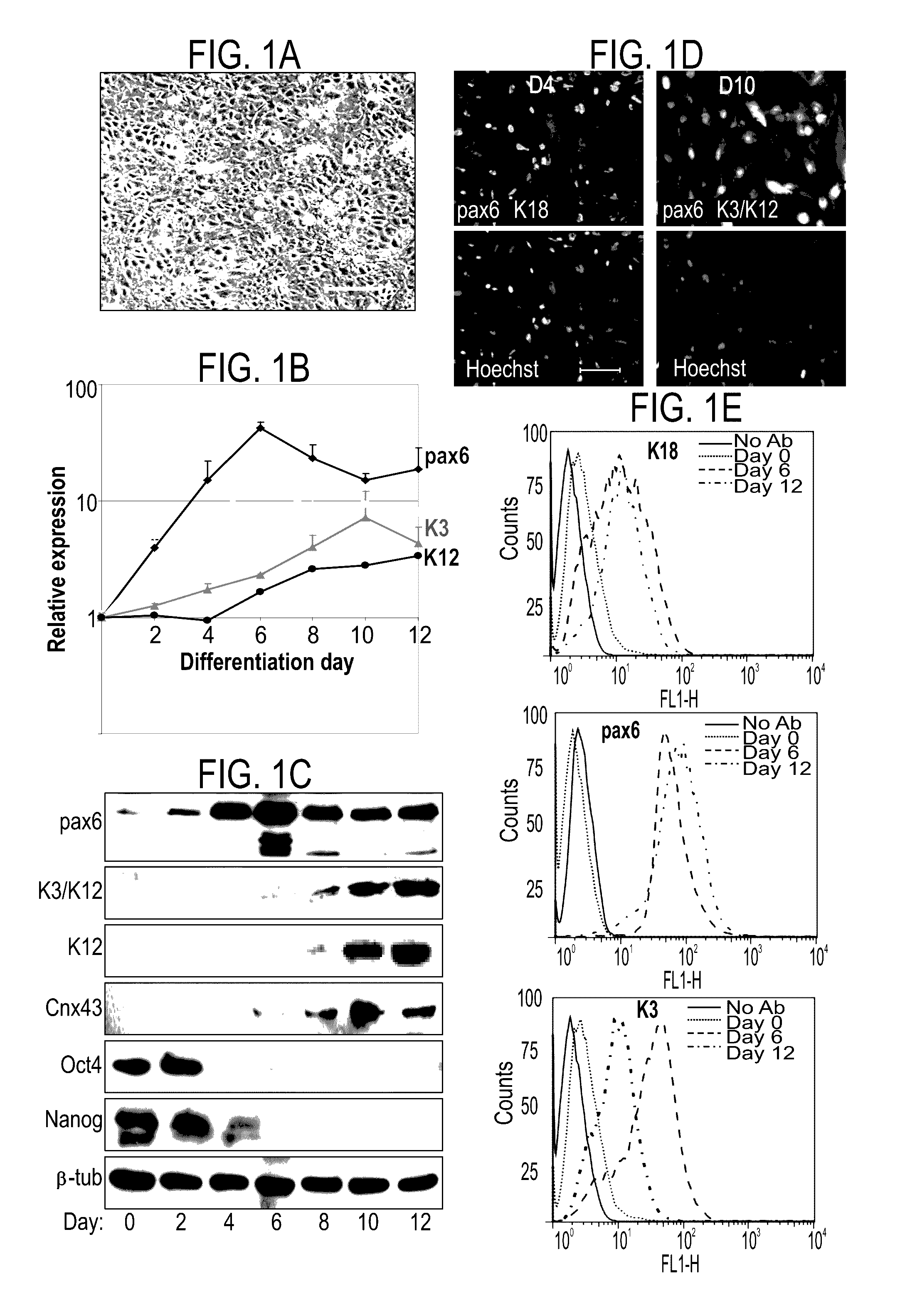
[0178]Real-time PCR analysis: RNA extractions of cells during different time point of corneal differentiation were analyzed by real-time PCR analysis of K3, K14, K12, K18, pax6 and DNp63.

[0179]FACS analysis: FACS analysis was performed using anti K14 Ab, anti K3 / K12 Ab and anti pax6 Ab.

[0180]Results

[0181]Addition of BMP-4 during the first three days significantly enhanced corneal differentiatio...


PUM

PropertyMeasurementUnit
densityaaaaaaaaaa
thicknessaaaaaaaaaa
sizeaaaaaaaaaa

Abstract

A method of generating a population of corneal epithelial cells is disclosed. The method comprises culturing human pluripotent stem cells in corneal fibroblast-conditioned medium on a solid surface comprising an extracellular matrix component thereby generating the population of corneal epithelial cells. Isolated cell populations and corneal tissues are also disclosed, as well as uses thereof.

Description

RELATED APPLICATIONS[0001]This application is a National Phase of PCT Patent Application No. PCT / IL2011 / 000914 having International filing date of Dec. 1, 2011, which claims the benefit of priority under 35 USC §119(e) of U.S. Provisional Patent Application No. 61 / 418,915 filed on Dec. 2, 2010. The contents of the above applications are all incorporated by reference as if fully set forth herein in their entirety.SEQUENCE LISTING STATEMENT[0002]The ASCII file, entitled 56481SequenceListing.txt, created on Apr. 29, 2013 comprising 1,276 bytes, submitted concurrently with the filing of this application is incorporated herein by reference.FIELD AND BACKGROUND OF THE INVENTION[0003]The present invention, in some embodiments thereof, relates to methods of generating corneal cells from pluripotent stem cells and cell populations comprising same.[0004]The cornea is a unique, transparent structure that covers the iris, pupil, and anterior chamber, providing most of the eye's optical power. T...

Claims


Application Information

Patent Timeline
Patent Type & Authority Patents(United States)
IPC IPC(8): C12N5/00C12N5/079G01N33/567C07K1/00C12N5/02G01N33/53
CPCC12N5/0062C12N5/0621C12N2501/11C12N2501/155C12N2501/33C12N2502/085C12N2502/1323C12N2506/02C12N2506/45C12N2533/54C12N2533/90G01N33/53G01N33/5023G01N33/5026G01N33/5073A61P27/02A61K35/30G01N33/5058C12N2533/52
Inventor ITSKOVITZ-ELDOR, JOSEPHABERDAM, DANIELSHALOM-FEUERSTEIN, RUBY
Owner INST NAT DE LA SANTE & DE LA RECHERCHE MEDICALE (INSERM)
Why Eureka
Try Eureka