Method for observing microscopic structure of stainless steel
A microstructure and stainless steel technology, applied in the observation field of stainless steel microstructure, can solve the problems of complex adjustment, unreported application development, and difficult sample preparation
- Summary
- Abstract
- Description
- Claims
- Application Information
AI Technical Summary
Problems solved by technology
Method used
Image

Examples
Embodiment 1
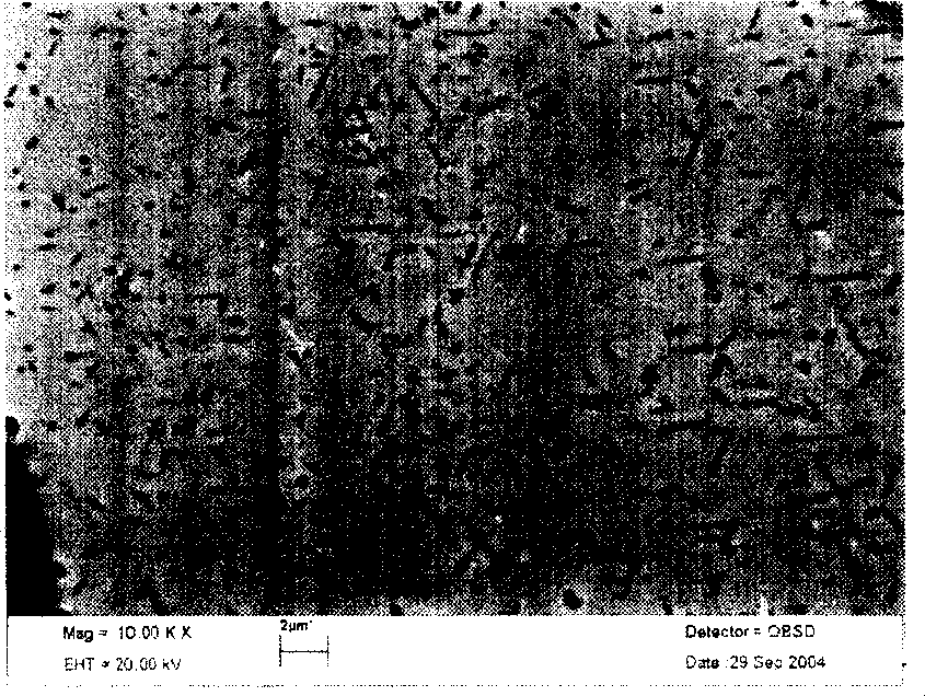
[0065] Example 1 Observation and Analysis of Dispersed Precipitated Phases in Stainless Steel
[0066] The observed stainless steel sample is 1Cr17Cu, and some fine and dispersed precipitates in stainless steel can only be observed by transmission electron microscope in the past. By adopting the microstructure observation method of the present invention, these precipitated phases can be clearly observed in a scanning electron microscope. In the development of antibacterial stainless steel, a very important link is the characterization and analysis of the microstructure of the sample after aging treatment, focusing on the analysis of whether there is ε-Cu phase precipitation, as well as the number, size and distribution of the precipitated phase. The observation method of the invention is used to observe and analyze the ε-Cu phase in 1Cr17Cu under different solid solution and aging systems, which can replace part of the analysis work of the transmission electron microscope. f...
Embodiment 2
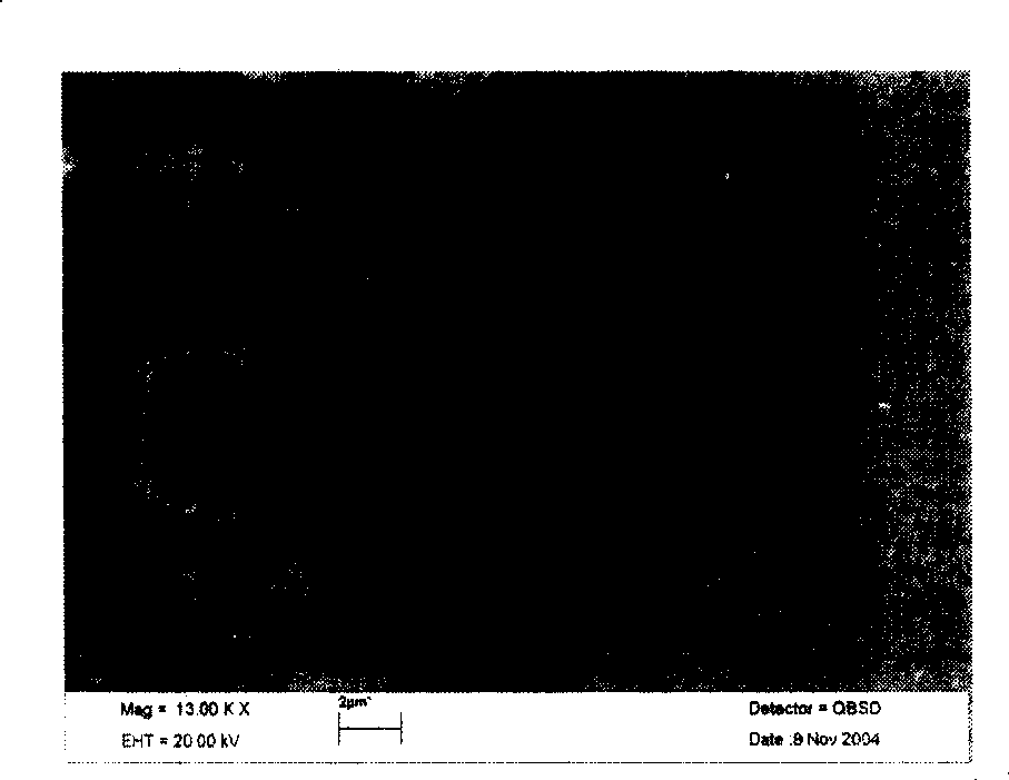
[0096] Example 2 Fine structure analysis of composite zone of stainless steel clad plate
[0097] What is observed is the composite interface of ferritic stainless steel / carbon steel (0Cr13Al / 16MnR) clad plate. Since the corrosion resistance of stainless steel and carbon steel is very different, when observing the structure by metallographic erosion, generally only one side can be eroded. Microstructure, or stainless steel and carbon steel are corroded separately, which has a great influence on the microstructure observation of the bonding interface, and it is difficult to observe clear microstructure features. This embodiment is a contrast image of backscattered electron crystal orientation after polishing of 0Cr13Al / 16MnR. Figure 4 It is a photo of the structure of the bonding area observed by the backscattered electron detector under the scanning electron microscope after the ferritic stainless steel 0Cr13Al and 16MnR are subjected to explosive compounding, annealing heat ...
Embodiment 3
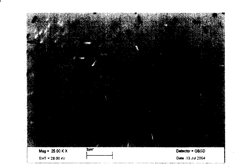
[0121] Example 3 Observation of grain boundary carbides after stainless steel sensitization treatment
[0122] The observed is 1Cr17Mn6Ni5N stainless steel. The carbide precipitated at the grain boundary of stainless steel during use is the main cause of intergranular corrosion. To test the performance of intergranular corrosion, the sample should be sensitized, and the precipitated carbide should be metallographically etched. It is difficult to identify the method, because the carbide will fall off after the grain boundary is eroded. By adopting the scanning electron microscope observation method of the present invention, the precipitated carbides can be visually observed. Figure 5 It is the grain boundary carbide morphology observed in sensitized 1Cr17Mn6Ni5N stainless steel.
[0123] The steps are as follows:
[0124] 1, the preparation of electrolytic polishing liquid is the same as embodiment one
[0125] 2. Sample processing and preparation Same as Example 1
[0126...
PUM
 Abstract
Description
Claims
Application Information
 - Generate Ideas
- Intellectual Property
- Life Sciences
- Materials
- Tech Scout
- Unparalleled Data Quality
- Higher Quality Content
- 60% Fewer Hallucinations
Browse by: Latest US Patents, China's latest patents, Technical Efficacy Thesaurus, Application Domain, Technology Topic, Popular Technical Reports.
© 2025 PatSnap. All rights reserved.Legal|Privacy policy|Modern Slavery Act Transparency Statement|Sitemap|About US| Contact US: help@patsnap.com