
Method for determining human plasma antiviral drug concentration

A technology of antiviral drugs and human plasma, applied in the field of medical testing, can solve the problems of being unsuitable for routine therapeutic drug concentration monitoring, cumbersome and time-consuming operations, and high analysis costs, achieving low cost, improved detection sensitivity, and less plasma consumption.

Inactive Publication Date: 2008-01-16
AFFILIATED HUSN HOSPITAL OF FUDAN UNIV
View PDF0 Cites 7 Cited by
  • Summary
  • Abstract
  • Description
  • Claims
  • Application Information

AI Technical Summary

Problems solved by technology

[0003] At present, in medical testing, there are domestic reports on methods for determining the concentration of relevant antiviral drugs in human plasma, but the method has various defects such as low sensitivity, cumbersome operation, time-consuming, low efficiency, and high analysis cost, and is not suitable for routine therapeutic drug concentration monitoring. ; There is no report on the simultaneous determination of the concentration of ACV, GCV and PCV in human plasma

Method used


Image

  • Method for determining human plasma antiviral drug concentration
  • Method for determining human plasma antiviral drug concentration

Examples


Embodiment 1

[0030] Chromatographic conditions:

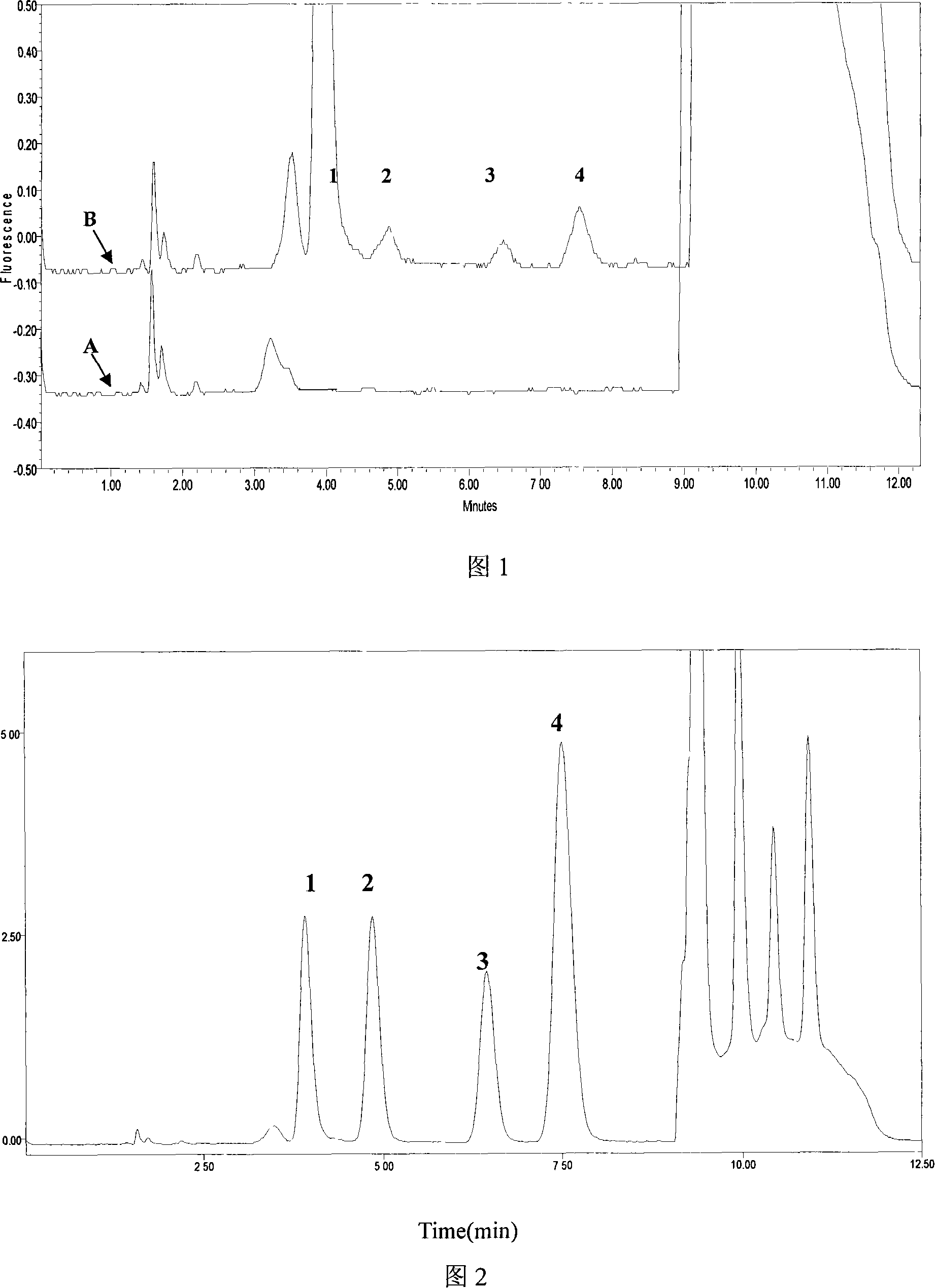
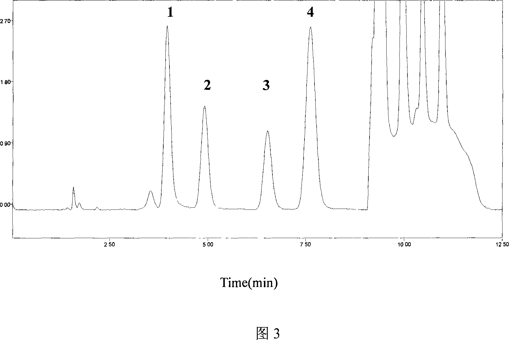
[0031] HPLC system: column Diamonsil C 18 (200mm×4.6mm, 5μm), the mobile phase adopts gradient elution, 0-7min is 0.08% TFA-methanol (96:4, V / V), 7.01-10.0min is 0.08% TFA-methanol (40:60, V / V), 0.08% TFA-methanol (96:4, V / V), 10.01~12.5min, flow rate 1.5mL min -1 ; Column temperature 25°C; fluorescence excitation wavelength 260±1nm, emission wavelength 380±1nm.

[0032] Plasma sample pretreatment:

[0033] Precisely draw 200 μL of plasma into a 1.5 mL centrifuge tube, add 50 μL of 7% perchloric acid solution containing internal standard guanylic acid, extract by vortexing for 30 seconds, centrifuge for 15 minutes (10000×g, 4°C), and take 40 μL of the supernatant into the The internal standard method was used to quantify the peak area.

[0034] Exclusiveness:

[0035] The blank plasma of 10 subjects who did not take ACV, GCV and PCV was taken from different sources, and measured according to the above sample pretreatment and measuremen...

Embodiment 2

[0043] Chromatographic conditions:

[0044] HPLC system: column Diamonsil C 18 (200mm×4.6mm, 5μm), the mobile phase adopts gradient elution, 0-7min is 0.1% TFA-methanol (96:4, V / V), 7.01-10.0min is 0.1% TFA-methanol (40:60, V / V), 10.01~12.5min for 0.1% TFA-methanol (96:4, V / V), flow rate 1.5mL min -1 ; Column temperature 25°C; fluorescence excitation wavelength 260±1nm, emission wavelength 380±1nm.

[0045] Plasma sample pretreatment:

[0046] Precisely draw 200 μL of plasma into a 1.5 mL centrifuge tube, add 25 μL of 20% perchloric acid solution containing internal standard guanylic acid, vortex for 30 s, centrifuge for 10 min (12000×g, 4°C), take 40 μL of supernatant and inject , the internal standard method was quantified by the peak area.

[0047] Exclusiveness:

[0048] The blank plasma of 10 subjects who did not take ACV, GCV and PCV was taken from different sources, and measured according to the above sample pretreatment and measurement methods, no interference of ...


PUM


Abstract

The invention belongs to medical detection field, relates to an analysis detection method of drug in the body of a person, and specifically relates to the method that the densities of antiviral drugs in blood plasma of the person such as acyclovir, ganciclovir and penciclovir can be detected at the same time. The method in the invention is characterized in that pilot sample is pretreated; as acyclovir, ganciclovir and penciclovir have the character of strong fluorescence absorption, acyclovir, ganciclovir and penciclovir can be separated from each other in an acidity flowing phase chromatographic column and be detected by a fluorescence detector. The method in the invention has the advantages of little sample, simple, swift and sensitive pretreatment, short analysis period and low cost; furthermore, the invention doesn't need expensive equipment and reagent and is suitable for the detection of clinical conventional blood drug density of acyclovir, ganciclovir and penciclovir.

Description

technical field [0001] The invention belongs to the field of medical testing, and relates to an analysis and determination method of drugs in vivo, in particular to a method for measuring the concentration of antiviral drugs in human blood plasma. Background technique [0002] At present, there are several antiviral drugs clinically used for the treatment of viral diseases. Nucleoside antiviral drugs. Among them, ACV and GCV are the most important anti-CMV drugs. PCV mainly has strong inhibitory effect on herpes simplex virus types I and II, varicella zoster virus and hepatitis B virus. When PCV was combined with ACV or GCV, the clinical efficacy was significantly enhanced. Because the pharmacokinetics of the three drugs vary greatly among individuals, combined medication is common, and abnormal liver and kidney function will lead to changes in drug plasma concentration and drug efficacy, it is important to adjust the dosage by detecting the concentration of the above dru...

Claims


Application Information

Patent Timeline
IPC IPC(8): G01N30/02G01N30/06G01N30/74G01N21/64
Inventor 道毅俊焦正钟明康
Owner AFFILIATED HUSN HOSPITAL OF FUDAN UNIV
Learn More