Preparation and application of polymer-coated magnetic nanoparticle contrast agent

A magnetic nanoparticle and polymer technology, applied in the field of medical imaging diagnostics, can solve the problems of cumbersome preparation methods of superparamagnetic contrast agents, and achieve the effect of safe use, good effect, and improved imaging contrast.

Inactive Publication Date: 2009-07-08
SHANGHAI NORMAL UNIVERSITY
View PDF0 Cites 8 Cited by
  • Summary
  • Abstract
  • Description
  • Claims
  • Application Information

AI Technical Summary

Problems solved by technology

Because the preparation method of superparamagnetic contrast agent is cumbersome, it is very necessary to invent a preparation method of superparamagnetic iron oxide polymer-wrapped magnetic nanoparticle contrast agent and its application in medical imaging

Method used


Image

  • Preparation and application of polymer-coated magnetic nanoparticle contrast agent
  • Preparation and application of polymer-coated magnetic nanoparticle contrast agent

Examples


Embodiment 1

[0039] Preparation of dextran-coated magnetic nanoparticles:

[0040] Get 3.22g ferric chloride (FeCl 3 ·6H 2 O), 1.18g ferrous chloride (FeCl 2 4H 2 O) and 2.5g dextran are dissolved in 100ml distilled water to form a transparent and stable microemulsion. Take 1ml of ammonia solution with a concentration of 28% and dilute it with 9ml of double distilled water, slowly add it to the microemulsion under constant stirring, and keep the temperature at 20°C for 130 minutes; centrifuge and precipitate to obtain stable dextran-wrapped magnetic nanoparticles Aqueous solution of particles;

[0041] The particle size is about 30-100nm as detected by transmission electron microscope (TEM).

Embodiment 2

[0043] Preparation of Magnetic Nano Contrast Agents:

[0044] 1. Take 2ml of dextran-coated magnetic nanoparticle aqueous solution, add 200ul10mol / L potassium periodate solution, and react in the dark for 5-8 hours;

[0045] 2. The above solution is centrifuged, and the separated magnetic nanoparticles are dissolved in a boric acid buffer solution with a pH of 8.0;

[0046] 3. Add 10-50ul mouse anti-human CA19-9 antibody to the boric acid buffer solution and react overnight at 4°C;

[0047] 4. Add potassium borocyanide solution to the above reacted boric acid buffer solution and react for 5-8 hours;

[0048] 5. The mixture is purified and separated, and then dispersed in a buffer solution with a pH of 7.4 to obtain a polymer-coated magnetic nanoparticle contrast agent.

Embodiment 3

[0050] In Vitro Magnetic Resonance Imaging of Pancreatic Cancer Cells Using Functional Magnetic Nanoparticles:

[0051] 1. Disperse 50ul polymer-coated magnetic nanoparticle contrast agent in cell culture medium (RPMI1640), and filter to sterilize;

[0052] 2. The particles obtained after filtration and the concentration are 1×10 6 Individual / mL pancreatic cancer cells BXPC-3 were co-incubated for 2 hours;

[0053]3. Digest with trypsin cell digestion solution (containing 0.25% trypsin and 0.02% ethylenediaminetetraacetic acid), and centrifuge to separate undigested substances;

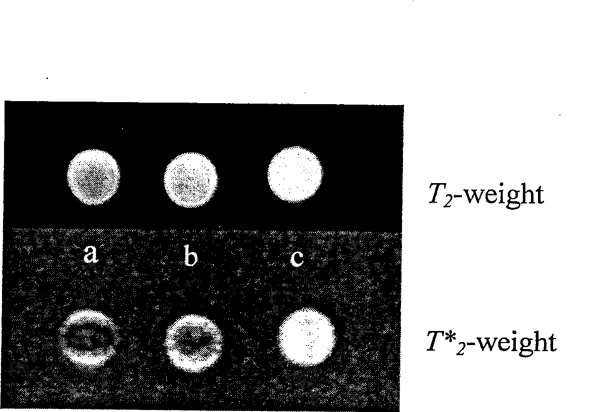
[0054] 4. Fix the above-mentioned treated cells with 0.5-2% glutaraldehyde, then disperse them in phosphate buffer solution, and perform magnetic resonance imaging in vitro. The results are shown in figure 1 .

[0055] figure 1 In a, the cell suspension with the magnetic contrast agent attached to the surface; b, the cell suspension; c, the PBS solution. It can be seen from the figure that compar...


PUM


Abstract

The invention relates to the field of medical image diagnosis and provides a preparation method and the application of macromolecule encapsulation magnetic nano-particle contrast agent. The current preparation method of superparamagnetic contrast agent is complex. The preparation of the macromolecule encapsulation magnetic nano-particle contrast agent comprises the steps as follows: iron chloride, iron dichloride and glucan are dissolved in secondary distilled water; strong ammonia is added slowly at the temperature of 15 DEG C to 40 DEG C to be constantly stirred for 130 to 150 minutes; centrifugal separation and deposition are carried out; potassium periodate solution is added; the processed solution is put into borate buffer solution with pH of 8.0; mouse anti-human CA19-9 antibody is added to be reacted at the temperature of 4 DEG C over a night; potassium borohydride is added to be reacted for 5 to 8 hours; and the mixture is purified and separated to be dispersed in buffer solution with pH of 7.4 to obtain the macromolecule encapsulation magnetic nano-particle contrast agent. The invention has the advantages that the invention improves the tumor magnetic resonance imaging contrast with good effect, has simple preparation method and is safe and convenient for use.

Description

technical field [0001] The invention relates to the field of medical imaging diagnostics, in particular to the preparation and application of a polymer-wrapped magnetic nanoparticle contrast agent. Background technique [0002] Contemporary Magnetic Resonance Imaging (MRI) technology is developing rapidly and becoming increasingly mature and perfect. It has been widely used in the field of biomedicine and has become an indispensable and important tool in clinical diagnosis and basic research. Magnetic resonance imaging is a new, non-invasive imaging method, which belongs to biological spin imaging technology, without the influence of radiation. Magnetic resonance images have high spatial resolution and very clear anatomical structure relationships, providing much more information than other imaging techniques in medical imaging. Moreover, the information provided is also different from the existing imaging techniques. The gray scale of the image reflects the strength of the...

Claims


Application Information

Patent Timeline
Patent Type & Authority Applications(China)
IPC IPC(8): A61K49/18A61K49/12A61K49/06
Inventor 沈鹤柏高明远黄明鸣缪飞
Owner SHANGHAI NORMAL UNIVERSITY
Why Eureka
Try Eureka