High-sensitivity high-flux DNA binding protein detection method

A technology of binding protein and detection method, applied in the field of DNA binding protein detection, can solve the problems of insufficient detection sensitivity, difficulty in detection of trace samples, increase in detection cost, etc., and achieve the effects of low cost, simple preparation, and reduced test cost.

Inactive Publication Date: 2009-12-02
XUZHOU MEDICAL COLLEGE
View PDF0 Cites 13 Cited by
  • Summary
  • Abstract
  • Description
  • Claims
  • Application Information

AI Technical Summary

Problems solved by technology

However, in the previous reported methods, on the one hand, different fluorescent probes were used to detect different proteins, which led to an increase in detection costs;

Method used


Image

  • High-sensitivity high-flux DNA binding protein detection method
  • High-sensitivity high-flux DNA binding protein detection method

Examples


Embodiment 1

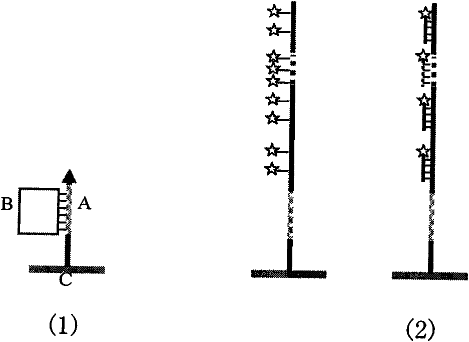
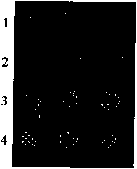
[0017] Achieve the detection of NF-kappaB p50, NF-kappaB p52 and OCT-1 proteins in the crude tumor cell culture extracts of the same cancer pain-sensitive sample: it will be used to detect NF-kappaB p50, NF-kappaBp52 and OCT-1 proteins, respectively The double-stranded oligonucleotide with acrylamide modification is fixed on the polyacrylamide gel chip (with wild-type sequence and known NF-kappaB p50 sample as positive control, with mutant sequence and known NF-kappaB p50 sample As a negative control), the crude tumor cell culture extract was incubated with the chip; then the double-stranded oligonucleotide on the chip was digested with exonuclease III. After the gel chip is decontaminated by low-frequency ultrasound, the loop detection probe is hybridized with the chip, and isothermal rolling circle amplification is performed for 30 minutes. Finally, the amplification chip is hybridized with the universal fluorescent detection probe, and the data is analyzed after the chip is sca...

Embodiment 2

[0019] Achieve the detection of NF-kappaB p50, NF-kappaB p52 and OCT-1 proteins in the crude tumor cell culture extracts of the same cancer pain-sensitive sample: it will be used to detect NF-kappaB p50, NF-kappaBp52 and OCT-1 proteins, respectively The double-stranded oligonucleotide with amino modification is fixed to the aldehyde modified chip (with wild-type sequence and known NF-kappaB p50 sample as positive control, and mutant sequence and known NF-kappaBp50 sample as negative Control), the crude tumor cell culture extract was incubated with the chip; then the double-stranded oligonucleotide on the chip was digested with exonuclease III. After the gel chip is decontaminated by low-frequency ultrasound, the loop detection probe is hybridized with the chip, and isothermal rolling circle amplification is performed for 30 minutes. Finally, the amplification chip is hybridized with the universal fluorescent detection probe, and the data is analyzed after the chip is scanned. The ...

Embodiment 3

[0021] Achieve the detection of NF-kappaB p50, NF-kappaB p52 and OCT-1 proteins in the crude tumor cell culture extracts of the same cancer pain-sensitive sample: it will be used to detect NF-kappaB p50, NF-kappaBp52 and OCT-1 proteins, respectively The double strands of biotin-modified oligonucleotides are fixed to streptavidin-modified magnetic microspheres or organic microspheres (with wild-type sequence and known NF-kappaB p50 samples as positive controls, and mutant type The sequence and the known NF-kappaB p50 sample are used as a negative control), the crude tumor cell culture extract is incubated with the chip; then the double-stranded oligonucleotide on the microsphere is digested with exonuclease III. After the microspheres are decontaminated by low-frequency ultrasound, the circular detection probe is hybridized with the chip, and isothermal rolling circle amplification is performed for 30 minutes. Finally, the amplified microspheres are hybridized with universal fluore...


PUM


Abstract

The invention belongs to microarray chip based DNA binding protein detection technology, and in particular relates to high-sensitivity high-flux DNA binding protein detection technology by a microarray chip. Double-stranded or single-stranded oligonucleotides are fixed on the chip, and a cell crude extract to be screened and the chip are directly incubated; then exonuclease is put into the system for incubation; and finally, a circular probe is added into the system for rolling circle amplification. Specific lattice sequences of the chip capable of combining a target protein can be taken as an extension primer for the rolling circle amplification; and sequences not combined with the protein are digested by the exonuclease, and cannot perform the rolling circle amplification, and finally amplified products and universal fluorescent probes are hybridized. The existence of the DNA binding protein can be detected according to the existence of specific array dot signals on the chip; and the intensity of the lattice fluorescence can quantitatively detect the content of the target protein. The chip can realize the detection of the DNA binding protein with super sensitivity, low cost, high specificity and high flux, and can be widely applied to scientific research and clinical detection and diagnosis.

Description

Technical field [0001] The invention belongs to a DNA binding protein detection technology based on a microarray chip, and particularly relates to a DNA binding protein detection method with high sensitivity and high throughput. Background technique [0002] For various life activities, the specific binding of protein and DNA is a key step for many cells to exhibit various biological activities, including gene transcription regulation, translation, and DNA replication, recombination and repair. Therefore, the detection of DNA-binding proteins and the identification of specific binding sites are very important for exploring gene expression mechanisms and analyzing cell functions. Furthermore, probing this information helps to find out the cause of the developmental distortion of biological organisms. Recently, DNA binding proteins and binding site information have become potential labels and targets for many disease diagnosis and drug discovery. Early diagnosis of related transcri...

Claims


Application Information

Patent Timeline
Patent Type & Authority Applications(China)
IPC IPC(8): C12Q1/68G01N21/64
Inventor 潘志强张励才邵翠杰张成标
Owner XUZHOU MEDICAL COLLEGE
Why Eureka
Try Eureka