Apparatus for collecting early-stage embryo or early-stage oocyte of pig and collecting method

A technology of oocytes and embryos, applied in veterinary equipment, medical science, animal delivery, etc., can solve the problems of egg flushing failure, lower egg flushing rate, fallopian tube bleeding, etc., and achieve the effect of free operation and improved success rate

Inactive Publication Date: 2010-08-11
INST OF ANIMAL SCI & VETERINARY HUBEI ACADEMY OF AGRI SCI
View PDF5 Cites 5 Cited by
  • Summary
  • Abstract
  • Description
  • Claims
  • Application Information

AI Technical Summary

Problems solved by technology

[0008] 1. During the flushing process, when pushing the syringe, because the syringe and the needle are rigidly connected, the operator will shake slightly or the pig will be a little disturbed, and the needle will easily scratch, puncture or fall out of the fallopian tube, resulting in tubal bleeding or flushing failure.
[0009] 2. Since the needle is inserted into the fallopian tube from the end of the fallopian tube at the junction of the uterine tube, first, it is easy to bleed; second, it is easy to puncture the fallopian tube; third, if the first flushing fails, the needle cannot be inserted from the same position for the second time, and it is necessary to Re-choose a site to insert the needle, thereby increasing the wound of the fallopian tube, and it is not easy to find a suitable site
[0010] 3. Since the egg-collecting tube is straight, the position of the egg-collecting dish is limited by the narrowness of the surgical department. If there is a little carelessness when picking up the eggs, or the pig is a little disturbed, the egg-washing liquid will easily flow out of the egg-collecting dish. Or the egg flushing liquid in the egg collection dish is splashed, resulting in a decrease in the egg flushing rate or failure of the egg flushing

Method used


Image

  • Apparatus for collecting early-stage embryo or early-stage oocyte of pig and collecting method
  • Apparatus for collecting early-stage embryo or early-stage oocyte of pig and collecting method
  • Apparatus for collecting early-stage embryo or early-stage oocyte of pig and collecting method

Examples


Embodiment 1

[0029] Embryo Collection and Transplantation of Excellent Breeding Pigs of Embodiment 1

[0030] Use high-quality purebred sows as donors, use hybrid sows that cannot be used as recipients as recipients, and transplant the embryos of donor sows to recipients of hybrid sows, with the purpose of using hybrid sows to produce high-quality purebred pigs. The operation steps are as follows:

[0031]1. Superovulation and mating of donor sows

[0032] On the 15th to 18th day of the estrous cycle of the donor sow, intramuscularly inject pregnant horse serum gonadotropin PMSG1000IU, and inject chorionic gonadotropin HCG800IU 72 hours later for superovulation treatment. After estrus, the boars of this breed are used for breeding.

[0033] 2. Simultaneous estrus of recipient sows

[0034] On the 15th-18th day of the estrous cycle of the recipient sow, while the superovulation treatment of the donor sow, PMSG 800IU was injected intramuscularly, and HCG 600IU was injected 72 hours later....

Embodiment 2

[0054] The preparation of embodiment 2 transhuman serum albumin gene pig

[0055] 1. Preparation of human serum albumin gene

[0056] Cut the constructed human serum albumin gene plasmid into linearity, and dilute it with TE diluent to a 5 μg / ml gene solution for later use.

[0057] 2. Collection of Embryos

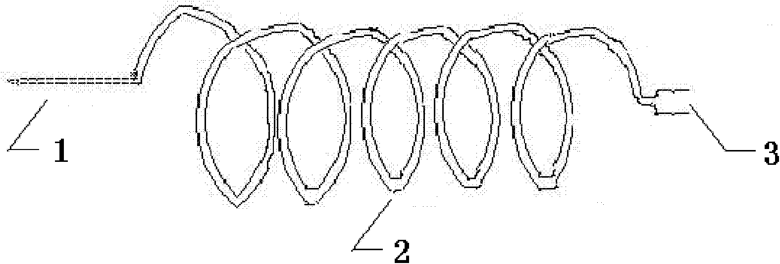
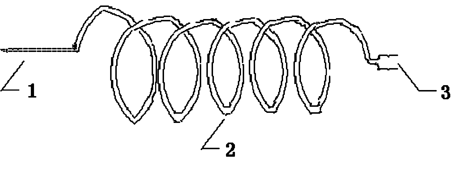
[0058] Using the same method as in Example 1, the sows were subjected to superovulation treatment and mated after estrus; 24 hours after the last mating, surgery, anesthesia, stabilization, and abdominal opening were carried out, and the process was the same as in Example 1. The operator extends the index finger and middle finger into the abdominal cavity through the incision, touches the uterus or uterine horn at the front and rear positions at the junction with the pelvic cavity, grasps it with two fingers, pulls it to the surface of the wound, and leads out the fallopian tube along one side of the uterine horn. Find the ovary at the end of the fallopian tube. First ...

Embodiment 3

[0070] The construction of embodiment 3 transfer EGFP gene clone pigs

[0071] 1. Preparation of donor cells

[0072] Take out the cryopreservation tube of transgenic porcine fetal fibroblasts from liquid nitrogen, put it in a 37°C water bath and shake carefully to make it thaw quickly; remove the cryopreservation solution by centrifugation, add 2mL culture medium with a pipette, and gently suspend cell. Adjust the cell density to 10 6 About cells / mL, serum-starved culture was carried out for 48-72 h in DMEM containing 0.5% fetal bovine serum. Afterwards, they were digested with trypsin and collected, and placed in the modified NCSU-23 phosphate culture medium for storage at 38.5°C for 20-120 minutes before being used for nuclear donors.

[0073] 2. Preparation of oocytes

[0074] Sows are not mated after estrus. Within 12 hours after the end of estrus, anesthesia and Baoding are used to make an incision, and the oviduct and ovary are drawn out of the incision; one end of ...


PUM


Abstract

The invention relates to an apparatus for collecting early-stage embryo or early-stage oocyte of a pig and a collecting method. The apparatus comprises an ovum collecting apparatus and an ovum collecting pipe, wherein the ovum collecting apparatus is a soft colloid pipe with the length of 200-300mm, the outer diameter of 1.0-1.1mm and the pipe wall thickness of 0.08-0.1mm, one end of the soft pipe is a No.9-12 pinhead and the other end is connected with a syringe; and the ovum collecting pipe has the length of 130-140mm, the wall thickness of 0.8-1.1mm and the inner diameter of 3-4mm with an angle of 120-140 degrees bending in the middle. When collecting early-stage embryo or early-stage oocyte of a pig, firstly, one end of the ovum collecting pipe is inserted into the fallopian tube for 10-15mm from fimbriae opening of the fallopian tube, and the other end is connected with an ovum collecting container; then the syringe sucked with 15-20ml of ovum collecting solution and 5-10ml of air is connected with the ovum collecting apparatus, the pinhead of the ovum collecting apparatus is inserted into the fallopian tube for 5-8mm from the uterine horn end of the junction of the uterine and the pipe, and then ovum collection is carried out. The invention obviously improves the success ratio and the efficiency of the collection.

Description

technical field [0001] The invention relates to a device and a collection method for collecting pig early embryos or oocytes, belonging to the field of animal embryo engineering. Background technique [0002] The collection and transplantation of early pig embryos was first reported by Kvashickii in 1951. By the 1960s, the technology of surgical method to collect and transfer embryos had been basically established. After the 1970s, North America and Europe began to apply the technology of collecting and transplanting embryos to the introduction of external blood and international introduction of closed pig herds. The emergence of transgenic animal technology in the 1980s has expanded the application of this technology. In 1985, Hammer et al. first collected fertilized eggs of pigs by means of surgery, then injected the human growth hormone gene into the pronuclei of fertilized eggs of pigs using microinjection technology, and then transferred it into the oviduct of sows to...

Claims


Application Information

Patent Timeline
Patent Type & Authority Applications(China)
IPC IPC(8): A61D1/00A61D19/00
Inventor 刘西梅李莉乔宪凤肖红卫魏庆信华文君熊忠良程妮华再东
Owner INST OF ANIMAL SCI & VETERINARY HUBEI ACADEMY OF AGRI SCI
Why Eureka
Try Eureka