Leukocyte classified counting reagent, kit and preparation method thereof and method of leukocyte classified counting

A white blood cell classification and kit technology, applied in biochemical equipment and methods, analysis materials, biological particle analysis, etc., can solve the problems of unclear classification, increase the complexity of instrument design, production cost, and unsatisfactory classification effect, and achieve Good production and application prospects, good classification effect, and short reaction time

Active Publication Date: 2011-03-23
SHENZHEN MINDRAY BIO MEDICAL ELECTRONICS CO LTD
View PDF8 Cites 26 Cited by
  • Summary
  • Abstract
  • Description
  • Claims
  • Application Information

AI Technical Summary

Problems solved by technology

[0010] In the above-mentioned prior art of using saponin non-ionic surfactants to classify leukocytes, most of them use two-step or three-step methods, requiring two or more reagents, which increases the complexity of instrumen

Method used


Image

  • Leukocyte classified counting reagent, kit and preparation method thereof and method of leukocyte classified counting
  • Leukocyte classified counting reagent, kit and preparation method thereof and method of leukocyte classified counting
  • Leukocyte classified counting reagent, kit and preparation method thereof and method of leukocyte classified counting

Examples


Embodiment 1

[0228] Fluorescent dye A 0.5ppm

[0229] Lauryl Glucoside 0.05g / L

[0230] Phosphate buffer 20mM

[0231] pH 7.0

[0232] Prepare 20nM phosphate buffer solution (pH 7.0), weigh the fluorescent dye and dodecyl glucoside according to the amount, add them to 800ml phosphate buffer solution, stir to dissolve, and dilute to volume with phosphate buffer solution. Filter and set aside.

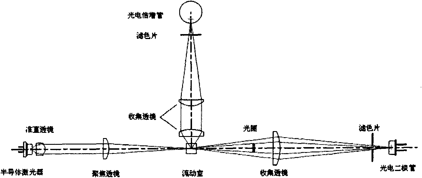
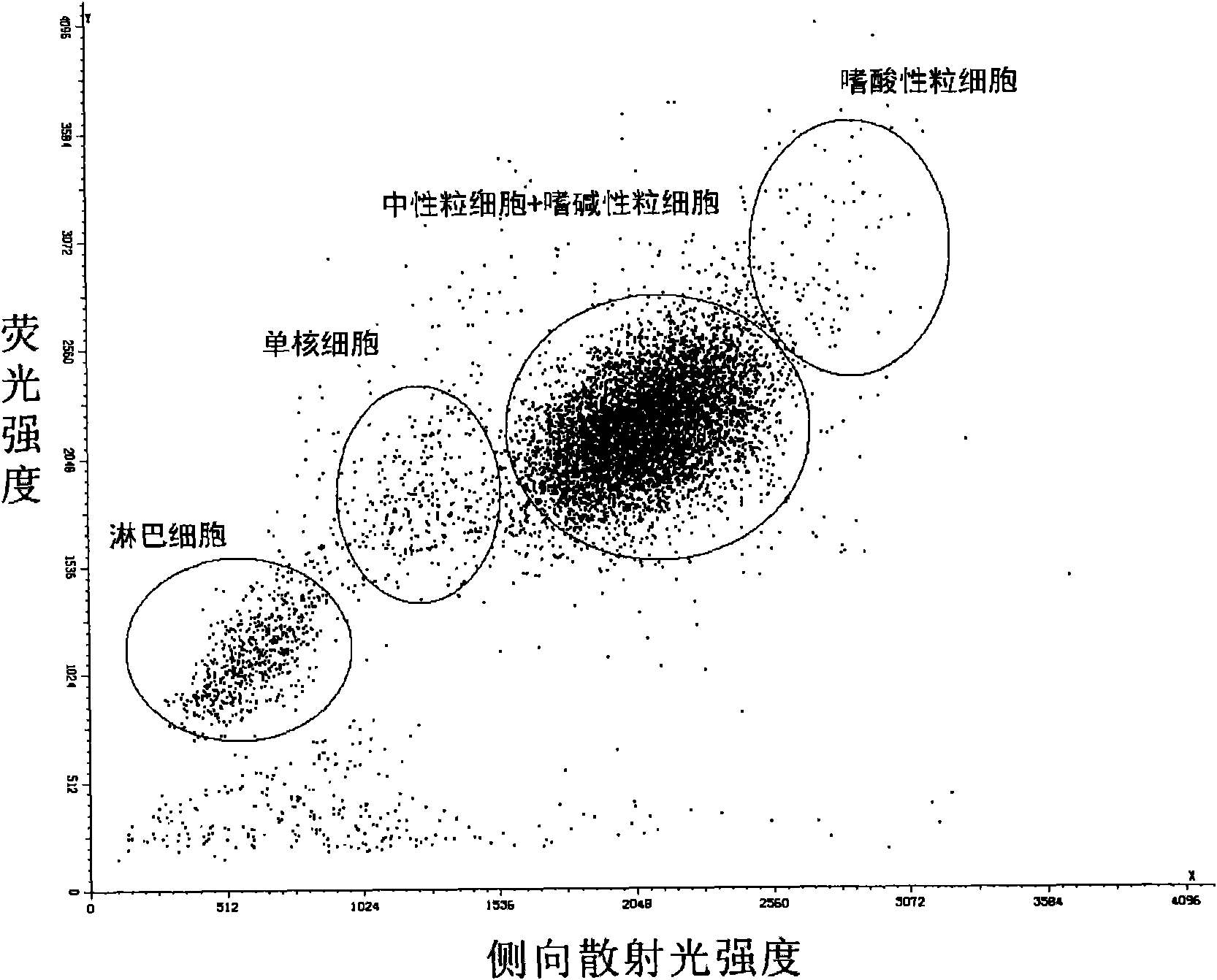
[0233] Add 20 μl of fresh anticoagulated blood to 1 ml of the above reagent, mix for 25 seconds while maintaining the temperature at 35° C., and then use laser flow cytometry (light source: red semiconductor laser, wavelength 633 nm) to detect leukocytes. The fluorescence intensity information of the cells is measured by using the side fluorescence at a measuring angle of 90 degrees, and the side scattered light intensity information of the cells is measured by using the side scattered light at a measuring angle of 90 degrees. The result is as figure 2 As shown, white blood cells are divided in...

Embodiment 2

[0235] Fluorescent dye E 0.5ppm

[0236] Saponin 0.05g / L

[0237] Polyoxyethylene (15) cetyl ether 0.8g / L

[0238] Phosphate buffer 20mM

[0239] pH 7.0

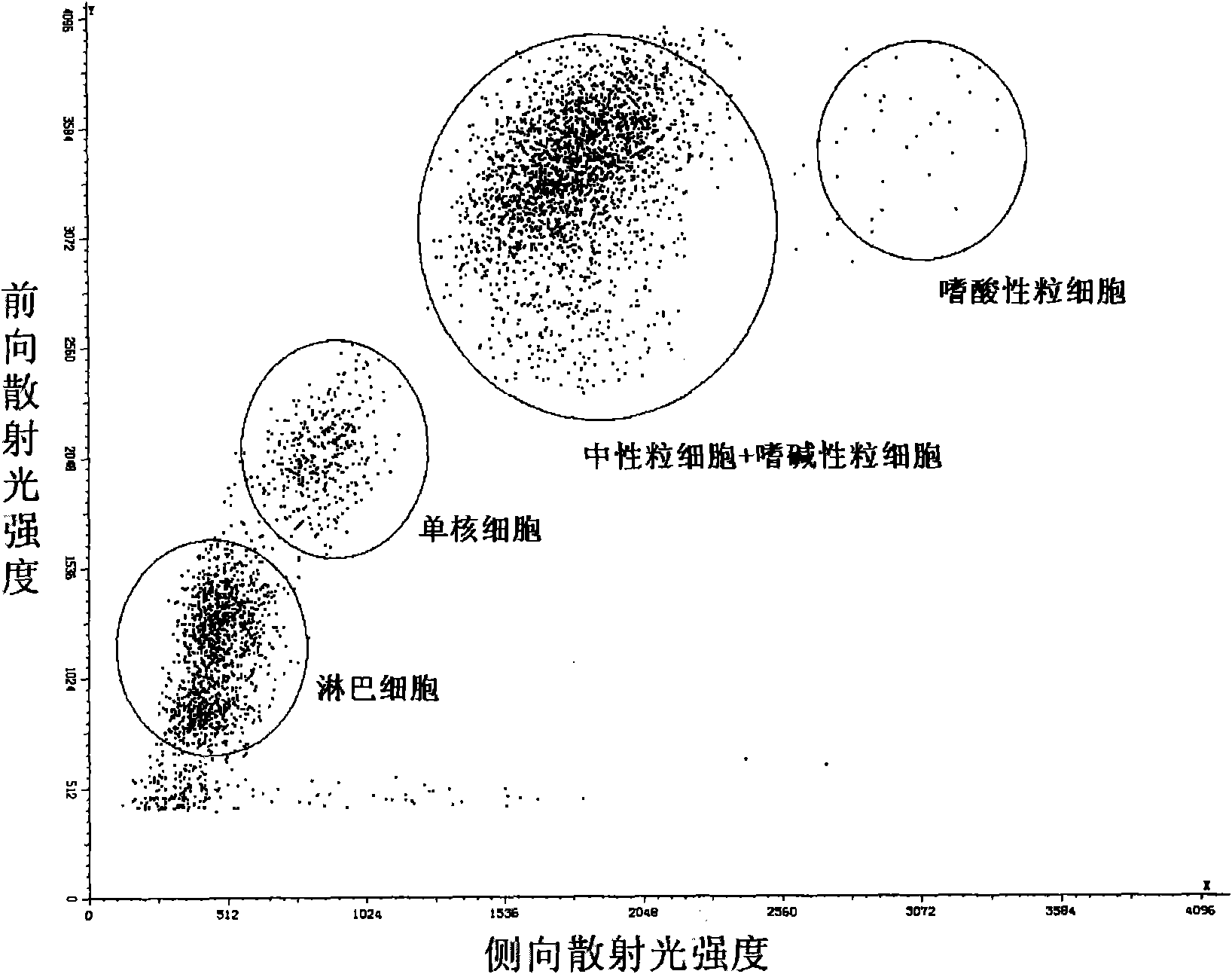
[0240] According to the same method as in Example 1, the above-mentioned components were used to prepare a solution, and the saponin here was purchased from Tokyo Chemical Industry Co., Ltd. Add 20 μl of fresh anticoagulated blood to 1 ml of the above reagent, mix for 23 seconds under the condition of maintaining the temperature at 43°C, and then use laser flow cytometry (light source: red semiconductor laser, wavelength 633nm) to detect leukocytes. The side scattered light intensity information of the cells is measured by using the side scattered light with a measuring angle of 90 degrees, and the forward scattered light intensity information of the cells is measured by using the low angle scattered light with a measuring angle of 1-5 degrees. The result is as image 3 As shown, white blood cells are divided into four ...

Embodiment 3

[0242] Fluorescent dye A 0.5ppm

[0243] Octyl Glucoside 0.1g / L

[0244] Sodium salicylate 2.0g / L

[0245] HEPES 20mM

[0246] Adjust pH 7.0 with NaOH

[0247] In the same manner as in Example 1, a solution was prepared using the above components. Add 20 μl of fresh anticoagulated blood to 1 ml of the above reagent, mix for 25 seconds while maintaining the temperature at 38°C, and then detect white blood cells by laser flow cytometry (light source: red semiconductor laser, wavelength 633nm). The fluorescence intensity information of the cells is measured by using the side fluorescence at a measuring angle of 90 degrees, and the side scattered light intensity information of the cells is measured by using the side scattered light at a measuring angle of 90 degrees. The result is as Figure 4 As shown, white blood cells are divided into four types: lymphocytes, monocytes, neutrophils and eosinophils.


PUM


Abstract

The invention discloses a leukocyte classified counting reagent, which includes: (1) cyanine type fluorescent dye; (2) glucoside compound. The invention further discloses a kit containing the leukocyte classified counting reagent and a preparation method thereof. The invention also discloses the reagent and a method f leukocyte classified counting for the kit. With the reagent, the kit and the method disclosed in the invention, the leukocyte can be counted in classification, wherein each subclass of the leukocyte has high discrimination index and good discrimination effect, and particularly, problems of difficulty in distinguishing a lymphocyte from a mononuclear cell and too short distance between an acidophilic cell and a neutrophile granulocyte are solved.

Description

technical field [0001] The present invention relates to blood cell classification and counting reagents and blood cell classification and counting methods, more specifically, the present invention relates to blood analysis reagents for classifying and counting white blood cells, a kit containing the reagents and a preparation method thereof, and using the The method for performing leukocyte differential counting with the above reagent or kit. Background technique [0002] Blood cells are roughly divided into three categories: red blood cells, white blood cells, and platelets, among which white blood cells are composed of five subtypes: lymphocytes, monocytes, neutrophils, eosinophils, and basophils. In the field of biological analysis, especially in the field of clinical test analysis, accurate classification of different subtypes of leukocytes plays a very important role in diagnosis and research, and the changes in the content of each subtype of leukocytes are used clinica...

Claims


Application Information

Patent Timeline
IPC IPC(8): C12Q1/06C12Q1/04G01N21/49G01N21/64
CPCG01N33/56972G01N2015/1486G01N2015/1477G01N15/147G01N2015/008
Inventor 赵阳雷霆赵玉梅徐兵
Owner SHENZHEN MINDRAY BIO MEDICAL ELECTRONICS CO LTD
Why Eureka
Try Eureka