Method for detecting mercury ion residue of fluorescent signal conversion mechanism based on nucleic acid aptamer structure

A nucleic acid aptamer, fluorescent signal technology, applied in fluorescence/phosphorescence, material excitation analysis, etc., to achieve the effect of small background value, less detection system and sample demand, and strong resistance to matrix interference

Inactive Publication Date: 2012-08-01
JIANGSU ACAD OF AGRI SCI
View PDF4 Cites 8 Cited by
  • Summary
  • Abstract
  • Description
  • Claims
  • Application Information

AI Technical Summary

Problems solved by technology

[0004]Use the DNA base sequence synthesized from the base sequence of the nickel ion RNA aptamer, and then modify some of its bases to obtain new mercury ions Nucleic acid aptamer, and design the complementary sequence Q2 matching with the mercury ion nucleic acid aptamer, establish a new fluorescent signal conversion mechanism, and use them to detect mercury ion residues, which has not been reported so far

Method used


Image

  • Method for detecting mercury ion residue of fluorescent signal conversion mechanism based on nucleic acid aptamer structure
  • Method for detecting mercury ion residue of fluorescent signal conversion mechanism based on nucleic acid aptamer structure
  • Method for detecting mercury ion residue of fluorescent signal conversion mechanism based on nucleic acid aptamer structure

Examples


Embodiment 1

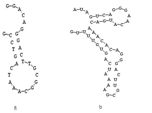
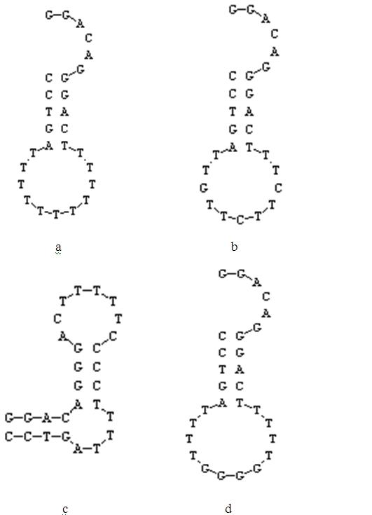
[0032] Construction of mercury ion nucleic acid aptamer N1

[0033] First, according to the base sequence of the nickel ion RNA aptamer reported in the literature, its DNA sequence was synthesized according to the method recorded in the literature Hofmann, HP, Limmer S, Hornung V, Sprinzl M. RNA, 1997, 3: 1289-1300, and its base The sequence is SEQ ID N O .7:

[0034] 5'-AUAGUCAGGGAACAUGACAAACACAGGGACUUGCGAAAAUCAGUGUUUUG-3'.

[0035] The base sequence is defined as DNA1-B.

[0036] When constructing the mercury ion nucleic acid aptamer N1, retain the 23rd to 44th bases of DNA1-B, and replace the 14th base A (equivalent to the 36th base of DNA1-B) with GC ( Add a base here), and finally add the base GG at the 5' end, and at the same time add the base CCG at the 3' end, and replace all the bases U in the reserved segment with the base T, that is, mercury Ionic nucleic acid aptamer N1, its base sequence is SEQ ID N O .2:

[0037] 5'-GGACAGGGACTTGCGGCAAATCAGTCCG-3'.

[0038...

Embodiment 2

[0040] Use the nucleic acid aptamer N1 to establish a fluorescent detection system:

[0041] Synthesize the complementary sequence Q2 with nucleic acid aptamer N1, its base sequence is SEQ ID N O .1:

[0042] 5'-GTCCCTGTCC-3';

[0043] Prepare bufferA buffer: 50mM NaCl+7mM MgCl 2 +50mM Tris / HCl, buffer pH=7.5;

[0044] Fluorescent group FAM, synthesized by Shanghai Jierui Biotechnology Co., Ltd., and it is labeled at the 5' end of the nucleic acid aptamer N1;

[0045] The fluorescence quenching group DABCYL was synthesized by Shanghai Jierui Biotechnology Co., Ltd., and it was labeled at the 3' end of the complementary sequence Q2.

[0046] The nucleic acid aptamer N1 labeled with the fluorescent group FAM and the complementary sequence Q2 labeled with the fluorescent quenching group DABCYL were respectively adjusted at a concentration ratio of 1:0~4 (the final concentration of the nucleic acid aptamer N1 was 50nmol / L, and the complementary sequence Q2 The final concentra...

Embodiment 3

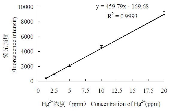
[0051] The detection standard curve of nucleic acid aptamer N1 was established by different concentrations of mercury ions.

[0052] The construction method of the fluorescence detection system is the same as that in Example 2.

[0053] Mix N1 and Q2 in the buffer A buffer system at a ratio of 1:3 (the final concentration of N1 is 150nmol / L, and the final concentration of Q2 is 450nmol / L). Denature at 94°C for 5 minutes, incubate at 25°C for 30 minutes, take out 50 μl from the system, and measure its fluorescence intensity; then add 50 μl of mercury ion standard solution in sequence, and carry out 2-fold gradient dilution of mercury ion concentration from 160ppm to 0.156ppm, And an equal volume of bufferA buffer was used as a control. After 5 minutes, the fluorescence intensity of each well was measured under the excitation light wavelength of 495 nm and the emission light wavelength of 535 nm. Standard curve see figure 2 ,Depend on figure 2 It can be seen that the corre...


PUM

PropertyMeasurementUnit
correlation coefficientaaaaaaaaaa

Abstract

The invention relates to a method for detecting mercury ion residue of a fluorescent signal conversion mechanism based on a nucleic acid aptamer structure. In the method, a mercury ion nucleic acid aptamer marked with a fluorescein molecule, and a complementary sequence marked with a quenching element are utilized to form a fluorescent detection system, wherein the mercury ion nucleic acid aptamer is a stem loop structure composed of 27-28 bases and owns a stem formed by covalent binding of the bases GGAC and GTCC; and the base sequence of the complementary sequence Q2 is shown in SEQ ID NO.1. The method for detecting the mercury ion residue by the fluorescent detection system comprises the following steps of: adding mercury ions with a serial concentration and a complementary sequence Q2 competitive binding nucleic acid aptamer; establishing a standard curve according to the change of fluorescent signals and determining the lowest detection limit and linear range; and then, carrying out marking detection on a sample to be detected and judging the content of the mercury ions in the sample according to the standard curve. The method disclosed by the invention has the advantages of rapidness, simplicity, high sensitivity and selectivity and less sample quantity demand, and is applicable to the detection of the mercury ions in the actual sample.

Description

[0001] technical field [0002] The invention relates to the detection range of mercury ion concentration, in particular to a method for detecting residual mercury ions based on the fluorescent signal conversion mechanism of the nucleic acid aptamer structure. [0003] Background technique [0004] Mercury is one of the important culprits of heavy metal pollution in the environment. It is a chemical substance with severe biological toxicity. Due to its persistence, easy migration and high bioaccumulation, mercury has become the most concerned environmental pollution. One of the substances, especially dissolved Hg 2+ It has high chemical activity and stability, and is the main form of pollutants discharged into environmental water bodies. It is corrosive and carcinogenic to cells. Therefore, a non-polluting, simple, fast, high-sensitivity and high-specificity method has been developed for The detection of mercury ions has become necessary, and the development and research of...

Claims


Application Information

Patent Timeline
Patent Type & Authority Applications(China)
IPC IPC(8): G01N21/64
Inventor 刘贤金雷兆静张存政刘媛
Owner JIANGSU ACAD OF AGRI SCI
Why Eureka
Try Eureka