Preparation method of protein-grain-supported-in-beaded-fiber tissue engineering fiber support frame

A protein particle and tissue engineering technology, applied in medical science, prosthesis, etc., can solve the problems of unable to optimize the protein release curve, unable to overcome the problem of protein drug burst release, etc., to improve the in vitro release curve, low cost and simple method. Effect

Inactive Publication Date: 2013-02-06
DONGHUA UNIV
View PDF7 Cites 32 Cited by
  • Summary
  • Abstract
  • Description
  • Claims
  • Application Information

AI Technical Summary

Problems solved by technology

However, since the fibers prepared by the electrospinning method are mostly hundreds of nanometers in diameter, they are not capable of loading granular drugs with a particle size of micron, so the release curve of the protein cannot be optimized, especially the burst release of the loaded protein drug cannot be overcome. question

Method used


Image

  • Preparation method of protein-grain-supported-in-beaded-fiber tissue engineering fiber support frame
  • Preparation method of protein-grain-supported-in-beaded-fiber tissue engineering fiber support frame
  • Preparation method of protein-grain-supported-in-beaded-fiber tissue engineering fiber support frame

Examples


Embodiment 1

[0029] (1) Preparation of A solution: Dissolve polylactic acid-glycolic acid copolymer in a mixed solvent of 0.25 mL N,N-dimethylformamide and 0.75 mL tetrahydrofuran to obtain A solution with a concentration of 150 mg / mL.

[0030] (2) Preparation of solution B: add 15 mg of dextran particles loaded with bovine serum albumin with a particle size of 227-1575 nm to solution A, and stir with magnetic force at 2000 rpm for 60 min.

[0031] (3) Electrospinning: Add B solution into the syringe, set the spinning voltage to 20kV, the spinning speed to 1mL / h, and the spinning distance to 20cm, and perform electrospinning on the receiver (a metal plate covered with a layer of aluminum foil). Collect the fibrous membrane.

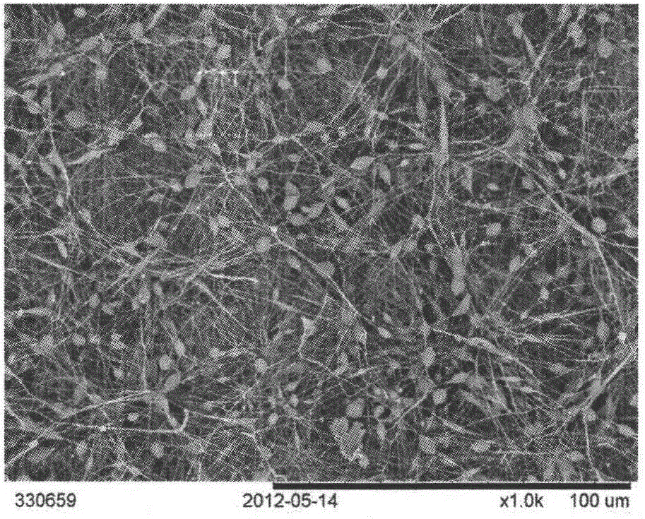
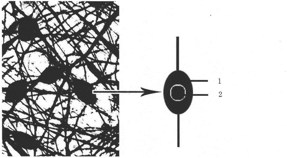
[0032] (4) The collected fiber membrane is left to dry at room temperature, and the receiver is peeled off to obtain the following: figure 1 A tissue engineering fibrous scaffold with beaded fibers loaded with protein particles is shown. Such as figure 2 As shown,...

Embodiment 2

[0036] (1) Preparation of A solution: Dissolve polylactic acid-glycolic acid copolymer in a mixed solvent of 0.25 mL N,N-dimethylformamide and 0.75 mL tetrahydrofuran to obtain A solution with a concentration of 150 mg / mL.

[0037] (2) Preparation of solution B: Add 10 mg of dextran particles loaded with bovine serum albumin with a particle size of 227-1575 nm to solution A, and stir with magnetic force at 2000 rpm for 60 min.

[0038] (3) Electrospinning: Add solution B into the syringe, spinning voltage 20kV, spinning speed 1mL / h, spinning distance 20cm, electrospinning, and collect fibers on the receiver (a metal plate covered with a layer of aluminum foil) membrane.

[0039] (4) The collected fiber membrane is left to dry at room temperature, and the receiver is peeled off to obtain a tissue engineering fiber scaffold with beaded fibers loaded with protein particles.

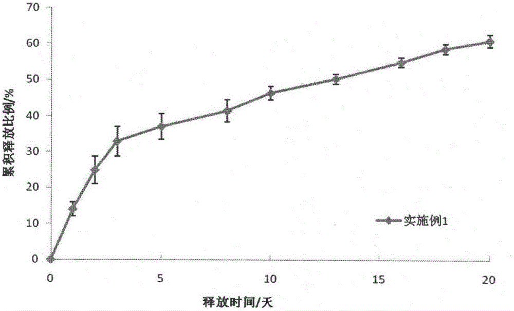
[0040] (5) Weigh 20 mg of fiber membrane, add 1 mL of PBS buffer solution with a pH of 7.4, and culture ...

Embodiment 3

[0043] (1) Preparation of A solution: Dissolve polylactic acid-glycolic acid copolymer in a mixed solvent of 0.25 mL N,N-dimethylformamide and 0.75 mL tetrahydrofuran to obtain A solution with a concentration of 150 mg / mL.

[0044] (2) Preparation of solution B: Add 6.67 mg of dextran particles loaded with bovine serum albumin with a particle size of 227-1575 nm to solution A, and stir with magnetic force at 2000 rpm for 60 min.

[0045] (3) Electrospinning: Add B solution into the syringe, set the spinning voltage to 20kV, the spinning speed to 1mL / h, and the spinning distance to 20cm, and perform electrospinning on the receiver (a metal plate covered with a layer of aluminum foil). Collect the fibrous membrane.

[0046] (4) The collected fiber membrane is left to dry at room temperature, and the receiver is peeled off to obtain a tissue engineering fiber scaffold with beaded fibers loaded with protein particles.

[0047] (5) Weigh 20mg of fiber, add 1mL of PBS buffer soluti...


PUM

PropertyMeasurementUnit
particle diameteraaaaaaaaaa
molecular weightaaaaaaaaaa
particle diameteraaaaaaaaaa

Abstract

The invention provides a preparation method of a protein-grain-supported-in-beaded-fiber tissue engineering fiber support frame, which is characterized by comprising the following specific steps: step 1, preparation of solution A: dissolving a high polymer into a solvent to obtain solution A; step 2, preparation of solution B: adding the protein grain into the solution A and uniformly agitating under magnetic force to obtain solution B; step 3, electrostatic spinning: adding the solution B into an injector, setting the spinning voltage, the spinning speed and the spinning distance, carrying out electrostatic spinning and collecting a fibrous membrane on a receiver; and step 4, standing and airing the collected fibrous membrane at a room temperature and stripping the receiver to obtain the protein-grain-supported-in-beaded-fiber tissue engineering fiber support frame. The preparation method has the beneficial effect that in the process of electrostatic spinning, a granular protein grain drug package is embedded in a fiber bead, so that the in-vitro release performances of drugs are improved.

Description

technical field [0001] The invention relates to a method for preparing a tissue engineering fiber scaffold in the technical field of medicine, in particular to a method for preparing a tissue engineering fiber scaffold using electrospun fibers to string beads and load protein particles. Background technique [0002] Research on electrospun fiber drug-loading systems began in the early 21st century. In 2002, Kenawy et al. published a paper entitled Release of tetracycline hydrochloride from electrospun poly(ethylene-co-vinylacetate), poly(lactic acid), and a blend (see: Journal of Controlled Release, 2002, 81 (1): 57-64), in the same year, Zong et al published a paper titled Structure and process relationship of electrospun bioabsorbable nanofiber membranes (see: Polymer, 2002, 43 (16): 4403-4412) papers, all proposed the idea of ​​using electrospun fibers to load drugs. Among them, Kenawy carried out electrospinning with polylactic acid (PLA), polyethylene-vinyl acetate (PE...

Claims


Application Information

Patent Timeline
Patent Type & Authority Applications(China)
IPC IPC(8): A61L27/24A61L27/22A61L27/20A61L27/18A61L27/16A61L27/54
Inventor 丁辛隋晓田玲玲李庭晓胡崛李毓陵王璐胡吉永杨旭东
Owner DONGHUA UNIV
Why Eureka
Try Eureka