Load small interfering RNA nanoscale lipid microbubble ultrasonic contrast agent and preparation method

A technology of ultrasonic contrast agent and lipid microbubble, which is applied in the field of ultrasonic molecular imaging and biomedical engineering, can solve the problems of large diameter, ineffective combination, and inability to load gene fragments, etc., to achieve integration and good enhancement of ultrasonic imaging effect of ability

Inactive Publication Date: 2013-05-15
THE THIRD AFFILIATED HOSPITAL OF SUN YAT SEN UNIV
View PDF3 Cites 21 Cited by
  • Summary
  • Abstract
  • Description
  • Claims
  • Application Information

AI Technical Summary

Problems solved by technology

[0003] However, commercialized microbubbles have obvious shortcomings as gene delivery vehicles (especially tumor-targeted gene delivery vehicles): ①The diameter is too large, because the ultrasound contrast agents currently on the market are all micron-scale, which can only limit In the vascular system, it cannot reach the surrounding tumor cells through the tumor vascular endothel...

Method used


Image

  • Load small interfering RNA nanoscale lipid microbubble ultrasonic contrast agent and preparation method
  • Load small interfering RNA nanoscale lipid microbubble ultrasonic contrast agent and preparation method
  • Load small interfering RNA nanoscale lipid microbubble ultrasonic contrast agent and preparation method

Examples


Embodiment 1

[0035] S1. Preparation of nano-scale lipid microbubble ultrasound contrast agent (hereinafter referred to as nano-microbubble) with negatively charged surface:

[0036] Nano-microbubbles were prepared using phospholipids as raw materials by thin film-hydration method. The phospholipid components dipalmitoylphosphatidylcholine (DPPC), distearoylphosphatidylethanolamine (DSPE), dipalmitoylphosphatidic acid (DPPA) were dissolved in chloroform in parts by weight 18:1:1 and placed in diameter 9 cm petri dish, in the fume hood to wait for the chloroform to volatilize naturally to form a phospholipid film. After adding 4 ml of double-distilled water and hydrating at 37 °C for 1 hour, the lipid solution was transferred to a 50 ml centrifuge tube, sonicated for 5 minutes with an ultrasonic breaker, and at the same time, octafluoropropane gas was introduced to prepare lipid microbubbles. The microbubble liquid was left to stand for 10 minutes, and then centrifuged at 1000 rpm for 5 min...

Embodiment 2

[0044] Example 2 Diameter and Surface Potential of Intermediate and Final Products:

[0045] The diameters and surface potentials of the intermediate products (nanobubbles, siRNA micelles) and final products (siRNA nanobubbles) obtained in Example 1 were detected by dynamic light scattering. The results (result data expressed as mean ± standard error) show that the diameters of nanobubbles, siRNA micelles, and siRNA nanobubbles are 436.8±5.7 nm, 66.8±2.8 nm, and 476.0±6.1 nm, respectively; and the surface potential of the three -18.4±0.2 mV; 23.4±1.1 mV, 15.3±2.7 mV. The diameter and surface potential verified the polymerization process of preparing siRNA nano-microbubbles.

Embodiment 3

[0046] Example 3 Morphological structure detection of siRNA nano-microbubbles:

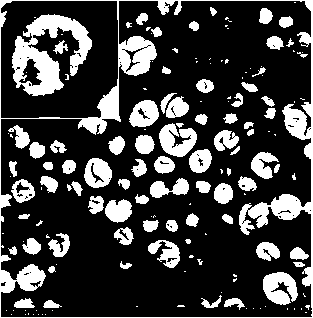
[0047] In order to further confirm the morphology and structure of the resulting siRNA nano-microbubbles prepared in Example 1, a transmission electron microscope is used to detect, and the test results are shown in figure 1 . Through the test, it can be seen that the siRNA nano-microbubbles are round or quasi-circular, and the diameter distribution is about 200-500 nm. The diameter distribution is uniform and there is no obvious aggregation. The microbubble surface dents caused by vacuuming during the preparation of the transmission electron microscope can be seen on the surface . Visible at high magnification ( figure 1 Upper left corner), the contrast agent is approximately round, the surface is not smooth, and a large number of small siRNA micelles with a diameter of about 50-70 nm can be seen on the surface of the microbubbles to form an overall structure.


PUM

PropertyMeasurementUnit
Diameteraaaaaaaaaa

Abstract

The invention discloses a load small interfering RNA (siRNA) nanoscale lipid microbubble ultrasonic contrast agent and a preparation method. The load siRNA lipid microbubble is formed by preparing DPPC (Dipalmitoyl Phosphatidyl Choline), DSPE (1, 2-distearoyl-sn-glycero-3-phosphoethanolamine) and DPPA (Diphenyl Phosphoryl Azide) into microbubbles (containing octafluoropropane) according to the weight part ratio of 18:1:1 and then assembling together with PEG-PLL (Polyethylene Glycol-Polylysne)-coated siRNA nanomicelle. The load siRNA nanoscale lipid is nanosclae, has an obvious ultrasonic contrast effect, and can generate obvious siRNA cell transfection efficiency under low-frequency ultrasonic irradiation, thereby further hopefully having important research values and application prospects in the fields of ultrasonic diagnosis and gene treatment.

Description

technical field [0001] The invention relates to the fields of ultrasound molecular imaging and biomedical engineering, in particular to a nanoscale lipid microbubble ultrasound contrast agent loaded with small interfering RNA (siRNA) and a preparation method thereof. Background technique [0002] As the third revolution in ultrasound imaging technology, contrast-enhanced ultrasound provides an effective basis for the diagnosis and differential diagnosis of various diseases, especially tumor diseases. With the rapid development of ultrasound imaging technology, it is found that microbubbles can be used as a contrast agent in ultrasound contrast on the one hand, and on the other hand can be used as the cavitation nucleus of ultrasound cavitation effect, which can reduce the threshold of cavitation effect and promote the targeting of gene fragments deliver. A large number of studies have proved that microbubbles as the core of cavitation can produce cavitation effect under low...

Claims


Application Information

Patent Timeline
IPC IPC(8): A61K49/22A61K48/00A61K47/34A61P35/00
Inventor 郑荣琴尹庭辉帅心涛
Owner THE THIRD AFFILIATED HOSPITAL OF SUN YAT SEN UNIV
Why Eureka
Try Eureka