Metastasis suppressor1 protein dimerization fluorescent probes and application thereof

A technique for metastatic tumors and fluorescent probes, applied in the field of fluorescent probe complementary proteins, to achieve the effects of simple processing, few steps and high accuracy

Inactive Publication Date: 2013-09-11
SOUTHEAST UNIV
View PDF2 Cites 1 Cited by
  • Summary
  • Abstract
  • Description
  • Claims
  • Application Information

AI Technical Summary

Problems solved by technology

I-BAR itself upregulates Rac activity, but does not induce ruffles, whic

Method used


Image

  • Metastasis suppressor1 protein dimerization fluorescent probes and application thereof
  • Metastasis suppressor1 protein dimerization fluorescent probes and application thereof
  • Metastasis suppressor1 protein dimerization fluorescent probes and application thereof

Examples


Embodiment 1

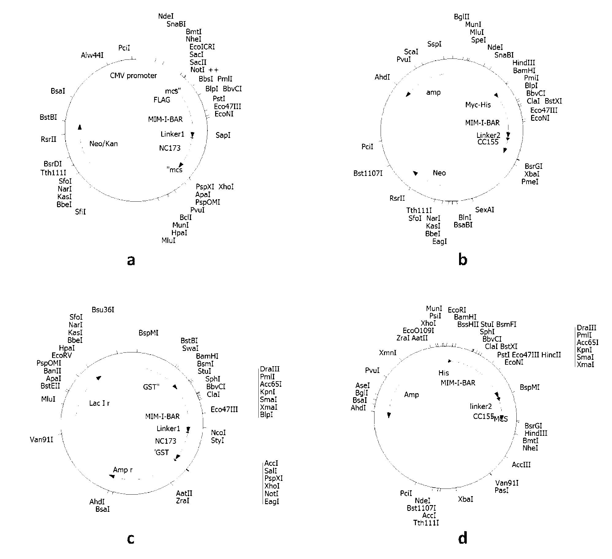
[0026] Example 1 Construction of plasmid DNA comprising polynucleotides represented by SEQ ID No: 1 and SEQ ID No: 3 respectively introducing Flag, Myc, GST and His tags.

[0027] (1) Construction of the N-terminal protein-encoding gene carrier of the fluorescent probe represented by Flag-labeled SEQ ID No: 2, that is, the plasmid (also known as pFlag-Mtss1-N) containing the polynucleotide represented by SEQ ID No: 1

[0028] The insert fragment was prepared using the overlap extension PCR method as follows: the DNA fragment Mtss1 (GenBankSEQ ID No: 211401) was used as a template, and the polynucleotides of 5'-CAGTTAGGATCCGAGGCTGTGTGATCGAG-3' and 5'-CGTAGCGATAGATCGGGGAGAAGAAGG-3' sequences were used as primers, Perform PCR amplification. The amplification reaction conditions are: 50μl reaction system, using PfuUltra TM II fusion HS DNA polymerase and its matching 10x buffer, 250 μM each of dNTP, 20 ng / μl template DNA, and 0.5 μM each primer. 30 cycles were performed on the M...

Embodiment 2

[0042] Example 2 Transfection and expression of fluorescent probe complementary proteins represented by SEQ ID No: 2 and 4 with Flag, Myc, GST, His tags, and corresponding polynucleotide recombinant plasmids in eukaryotic / prokaryotic cells.

[0043] Human embryonic kidney cell 293T cells were cultured using DMEM high-glucose medium containing 10% calf serum BCS, adding penicillin at a final concentration of 100 units / ml and streptomycin at a final concentration of 100 μg / ml. Before DNA transfection, 1.5x10 6 Cells were seeded in 100mm Petri dishes using normal medium without antibiotics at 37°C and 5% CO 2 conditions overnight. The transfection mixture was then prepared: 5 μg plasmid pFlag-Mtss1-N, 5 μg plasmid pMyc-Mtss1-C, 300 μl DMEM, 60 μl SuperFect. After the transfection mixture was incubated at room temperature for 5-10 minutes, add 3ml of normal culture medium and mix well to replace the cell culture medium and culture the cells for 2-3 hours, then replace it with no...

Embodiment 3

[0046] Example 3 The corresponding recombinant plasmid expression vector comprising the polynucleotide represented by SEQ ID No: 1 and 3, and the detection of the expression product.

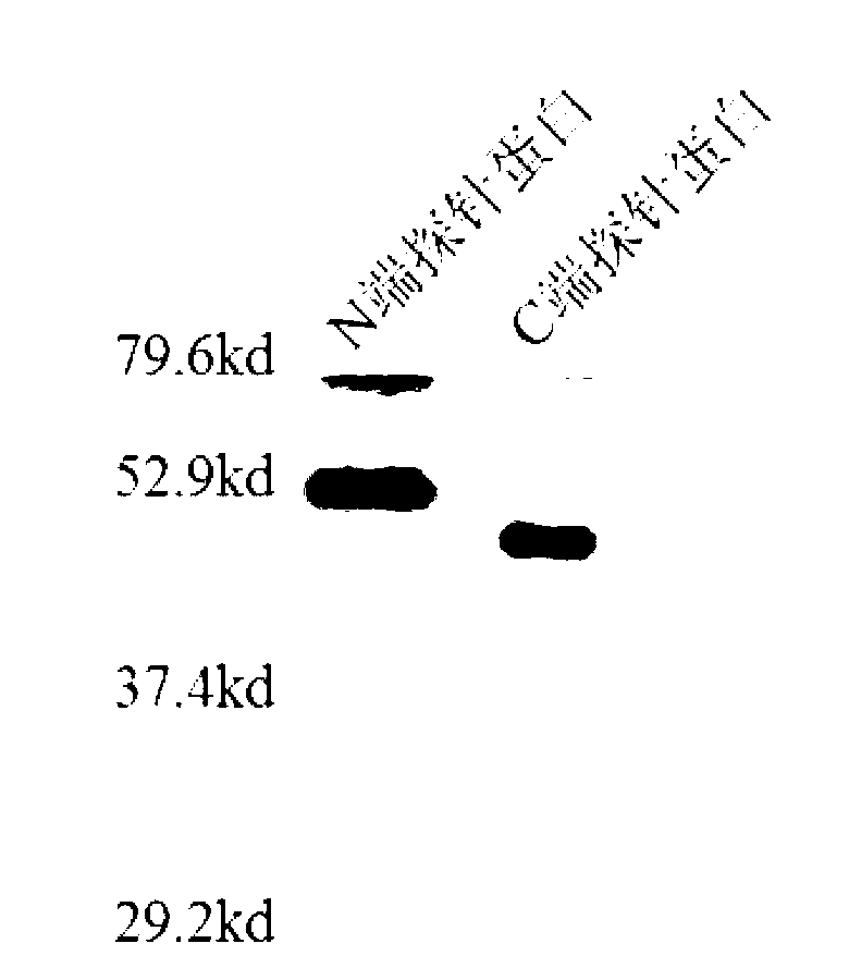
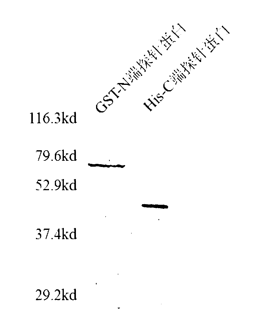
[0047] After eukaryotic or prokaryotic cells are transfected with recombinant plasmid expression vectors, the detection and evaluation methods of expression products mainly include Western Blot detection method, Coomassie brilliant blue detection method and immunofluorescence detection method.

[0048] Western Blot detection method is as follows. Chromatography of cell lysates or purified protein samples by 10% SDS-polyacrylamide gel electrophoresis. After the electrophoresis, wet electrotransfer the gel to the nitrocellulose membrane at 350mA current at 4°C for 1h, incubate overnight at 4°C with the 1:500-1:2000 dilution of 5% skimmed milk of the corresponding antibody, and buffer with TBST. After washing with horseradish peroxidase-labeled secondary antibody IgG1:200~1:1000, incubate at room te...


PUM


Abstract

The invention provides a group of metastasis suppressor1 (Mtss1) protein dimerization fluorescent probe complementary proteins containing amino acid sequences as shown in SEQ ID No: 2 and SEQ ID No: 4 which respectively correspond to a fluorescence probe N-terminal protein and a fluorescence probe C-terminal protein. The invention also provides polynucleotides encoding the group of proteins and recombinant vectors of the polynucleotides. The fluorescence probes provided by the invention can make use of dimerization of an Mtss1 protein, and end-connecting complementation genes of the fluorescence probes are close to each other and interact with each other to form an ability to excite fluorescence. The fluorescence intensity of the probes can indicate the dimerization extent of an I-BAR district of the Mtss1 protein, and shows a linear relationship with activities of Mtss1 protein I-BAR inhibiting drugs. The probe proteins provided by the invention can be used for studying an Mtss1 protein dimerization process, and can also be used for high efficiency screening of Mtss1 protein inhibitors.

Description

technical field [0001] The invention belongs to metastatic tumor deletion protein, in particular to a group of fluorescent probe complementary proteins for the dimerization of metastatic tumor deletion protein. Background technique [0002] BAR (Bin-Amphiphysin-Rvs) family proteins [McMahon, H.T.; Gallop, J.L. (2005) Nature 438, (7068), 590-6.] contain a highly conserved N-terminal domain in structure, often forming a The typical dimer structure is involved in the movement process of the cell membrane [Rao, Y.; Haucke, V. (2011) Cell Mol Life Sci68, (24), 3983-93.]. Metastasis suppressor1 (hereinafter referred to as Mtss1) protein belongs to the inverse BAR (I-BAR) subfamily of BAR superfamily proteins. The dimer of its N-segment domain looks like a distorted ellipsoid, and binds to the cell membrane through its convex surface. Other I-BAR proteins include insulin receptor tyrosine kinase substrate p53 (IRSp53), actin-binding protein with BAIAP2 homology region (ABBA), insu...

Claims


Application Information

Patent Timeline
IPC IPC(8): C07K14/00G01N33/68C12N15/63
Inventor 吉民常维维詹熙顾宁曹萌
Owner SOUTHEAST UNIV
Why Eureka
Try Eureka