Preparation and detection methods of screen-printed electrode immunosensor for rapidly detecting microcystin

A screen-printed electrode and immunosensor technology, which is applied in the field of screen-printed electrode immunosensors modified by composite magnetic nanomaterials, can solve problems such as methods that have not yet been published, and achieve the effects of low cost, simple and fast method, and high sensitivity

Active Publication Date: 2013-09-18
网都河北科技服务有限公司
View PDF7 Cites 19 Cited by
  • Summary
  • Abstract
  • Description
  • Claims
  • Application Information

AI Technical Summary

Problems solved by technology

Currently, core-shell Fe 3 o 4 Related technologies and methods for detectin

Method used


Image

  • Preparation and detection methods of screen-printed electrode immunosensor for rapidly detecting microcystin
  • Preparation and detection methods of screen-printed electrode immunosensor for rapidly detecting microcystin

Examples


Embodiment 1

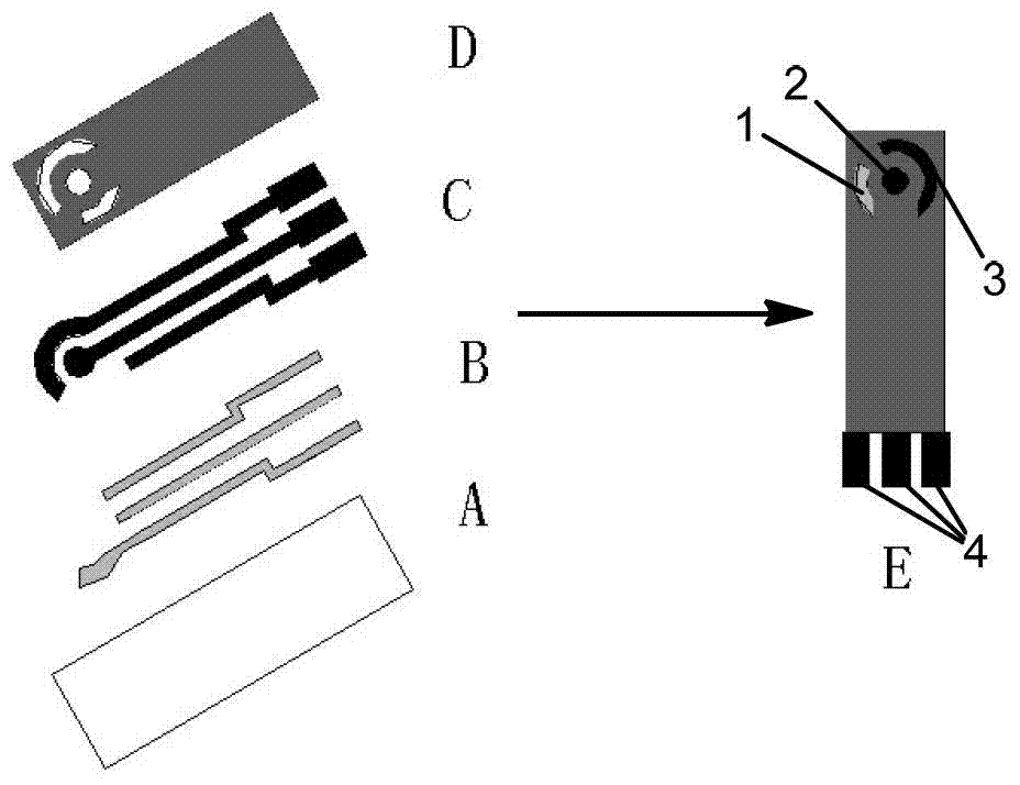
[0020] Structure and preparation process of screen-printed electrodes

[0021] see figure 1 , The screen printing electrode used in the present invention is to screen print three electrodes on the polyvinyl chloride film: one is a silver pseudo-reference electrode 1, one is a working electrode 2, and one is a counter electrode 3. The other end of the electrode is the lead terminal 4 of the three electrodes. On the substrates other than the three electrode lead terminals and the three electrodes, an insulating layer D printed with insulating paste is also covered.

[0022] Firstly, the silver paste was slowly stirred evenly, prepared by screen printing technology and printed separately on a polyvinyl chloride film with a thickness of 0.5mm ( figure 1 A), put it in an oven and dry it to make a semi-annular reference electrode and a conductive base rail ( figure 1 B); Then print the semi-annular carbon counter electrode, the circular working electrode and the lead wires of the...

Embodiment 3

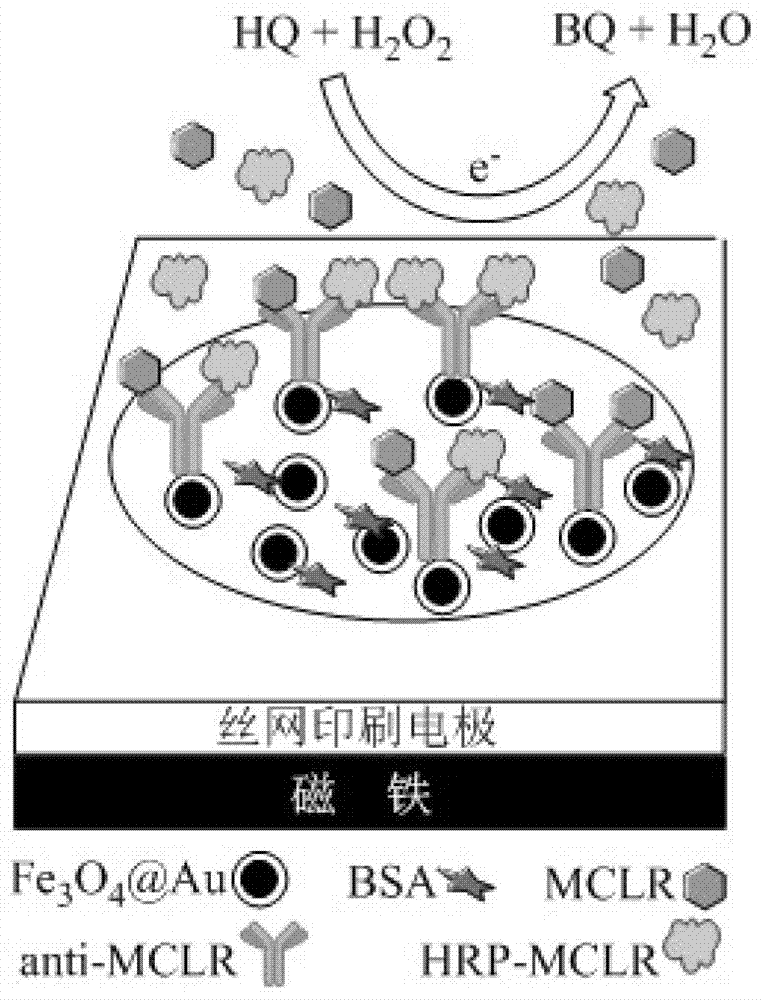
[0026] Embodiment 3: the method for detecting microcystin

[0027] When the microcystin electrochemical sensor of the present invention is applied, a potentiostat is required. First, add 10 μL of MCLR standard solutions of different concentrations and 10 μL of 10 mg / L MCLR-HRP dropwise onto the surface of the immunological working electrode, incubate at room temperature for 20 min, rinse with secondary water, and blow dry carefully with nitrogen; then, the reacted electrode Add 1mmol / L H 2 o 2 and 0.1mol / L PBS (pH7.0) of 1mmol / L HQ, reacted for 20s, and measured by differential pulse voltammetry (DPV), and obtained a standard curve of MCLR and oxidation peak current. Then the water samples were detected by the same method, and the concentration of MCLR in the water samples was obtained by comparing with the standard curve. The potential scanning range is -0.2V to 0.8V, the pulse amplitude is 50mV, and the pulse width is 50ms.


PUM


Abstract

The invention discloses preparation and detection methods of a screen-printed electrode immunosensor for rapidly detecting microcystin, and belongs to the field of environment monitoring technologies. Through a magnetic field formed by a magnet below a screen-printed electrode, core-shell magnetic nanoparticles Fe3O4@Au are fixed on the surface of a working electrode; through the adsorption between nano-Au and a microcystin antibody, the microcystin antibody is fixed on the surface of the electrode to prepare an MCLR antibody electrode; a certain concentration of MCLR and MCLR which is marked by horse radish peroxidase are together modified on the surface of the working electrode; after immunoreaction is performed for a period of time, a peak current value is measured by using DPV (differential pulse voltammetry) to obtain standard curves of the MCLR and the oxidation peak current; the steps are repeatedly carried out on water samples to be detected, and the obtained oxidation peak currents are compared with standard curves to obtain the MCLR concentration. According to the preparation and detection methods of the screen-printed electrode immunosensor for rapidly detecting microcystin, benefit is brought to rapidly detect the MCLR, the water samples do not need purification treatment, simplicity and rapidness are achieved, and the detection cost is reduced.

Description

technical field [0001] The invention belongs to the technical field of environmental monitoring, and in particular relates to a screen printing method modified by composite magnetic nanomaterials for detecting Microcystin-(leucine-arginine) (MCLR) Electrode immunosensor. Background technique [0002] Microcystins (MCs) is a heptapeptide monocyclic hepatotoxin with strong toxicity and great acute harm produced by cyanobacteria blooms. Its molecules have a ring structure and spacer double bonds, which are quite stable. . It has an inhibitory effect on protein phosphatase 1 and protein phosphatase 2A, and also has a strong hepatocarcinogenic effect, among which MCLR is one of the most common and most acutely toxic microcystins. As the degree of eutrophication in China's water bodies has gradually increased, the occurrence of cyanobacterial blooms has also gradually increased. 80% of the cyanobacteria blooms can detect the secondary metabolites—microcystins. Its harm to the w...

Claims


Application Information

Patent Timeline
IPC IPC(8): G01N33/553G01N27/327G01N27/48
Inventor 康天放张金果薛瑞
Owner 网都河北科技服务有限公司
Why Eureka
Try Eureka