

Contrast agent as well as preparation method and application thereof
A contrast agent and nanoparticle technology, which can be used in nuclear magnetic resonance/magnetic resonance imaging contrast agents, pharmaceutical formulations, preparations for in vivo tests, etc., can solve problems such as limited applications, and achieve good relaxation rates and fluorescence quantum yields. The effect of height and size is easy to control
- Summary
- Abstract
- Description
- Claims
- Application Information
AI Technical Summary
Problems solved by technology
Method used
Image

Examples
Embodiment 1
[0050] Preparation of contrast agent
[0051] 1) Dissolve 0.075mmol of manganese acetate and 0.3g of polyvinylpyrrolidone in a mixed system of 15mL of ethanol and 5mL of water to obtain solution A;
[0052] 2) Dissolve 0.04mmol potassium cobalt cyanate in 10mL water to obtain solution B;
[0053] 3) Use a syringe to slowly add solution B to solution A under magnetic stirring conditions, stir for 1 hour, then let the resulting cloudy liquid stand for 24 hours, then centrifuge, wash and dry to obtain Mn 3 [Co(CN) 6 ] 2 Nanoparticles;
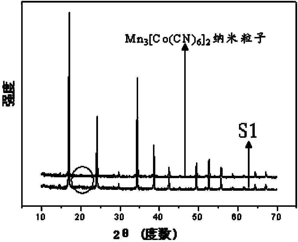
[0054] 4) Take 18mg Mn 3 [Co(CN) 6 ] 2 Nanoparticles were dispersed in 18mL of ethanol, added ammonia water and stirred for 10 minutes, then added 68μL tetraethyl orthosilicate, stirred for 2 hours, then centrifuged, washed and dried to obtain Mn 3 [Co(CN) 6 ] 2 Contrast agent S1 with nanoparticles as the core and silica coating as the shell.
[0055] Such as figure 1 As shown, S1 consists of Mn 3 [Co(CN) 6 ] 2 and silicon dioxide, a...
Embodiment 2
[0059] T of S1 1 -T 2 Dual Mode MRI Effects
[0060] Such as Figure 4 Shown, T 1 In the imaging mode, the image changes from dark to bright from low density to high density, T 2 In the imaging mode, it is just the opposite, showing the dual-mode imaging contrast effect of S1;
[0061] Such as Figure 5 As shown, S1 can continuously provide T at the mouse brain tumor site 1 and T 2 Contrast imaging effect;
[0062] Such as Figure 6 As shown, the curve of MRI signal decline with time in (b) shows that the brain T after S1 injection 2 The decrease of the signal, and by comparing the tumor tissue (pointed by 1 in (a)) and normal tissue (pointed by 2 in (a), it can be seen that due to the enrichment of S1 in the tumor site, its T 2 Significantly lower signal than normal tissue;
[0063] Such as Figure 7 As shown, S1 can accumulate in the kidney through the circulation in the body to provide continuous contrast of the kidney, and can provide excellent T in the blood v...
Embodiment 3
[0065] Fluorescence imaging effect of S1
[0066] Such as Figure 8 As shown, the phagocytosis of Mn 3 [Co(CN) 6 ] 2 Tumor cells after nanoparticles (a) and S1 (b) showed fluorescent signals under multiple bands. Among them, under the excitation wavelength of 403nm, the fluorescence signal presents a weak blue light, and under the excitation wavelength of 488nm, the fluorescence signal imaging presents a strong green light, while in two-photon excitation, the fluorescence signal presents a strong blue light under the excitation wavelength of 720nm , which demonstrates that S1 can provide usable fluorescence signals in multiple wavelength bands as well as in two-photon fluorescence imaging.
PUM
 Abstract
Description
Claims
Application Information