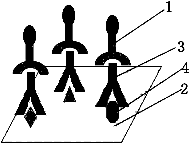
Systemic lupus erythematosus (SLE) autoantibody detector
A lupus erythematosus and autoantibody technology, applied in the field of biomedicine, can solve the problems of long diagnosis time, accuracy to be improved, high cost, etc., and achieve the effect of excellent specificity and excellent sensitivity
- Summary
- Abstract
- Description
- Claims
- Application Information
AI Technical Summary
Problems solved by technology
Method used
Image
Examples
Embodiment 1
[0026] (1) Chip production:
[0027] Step 1: Cut the cellulose acetate membrane with a 0.2 μm pore size to 25cm×75cm, and place it in the card slot of the spotting instrument.
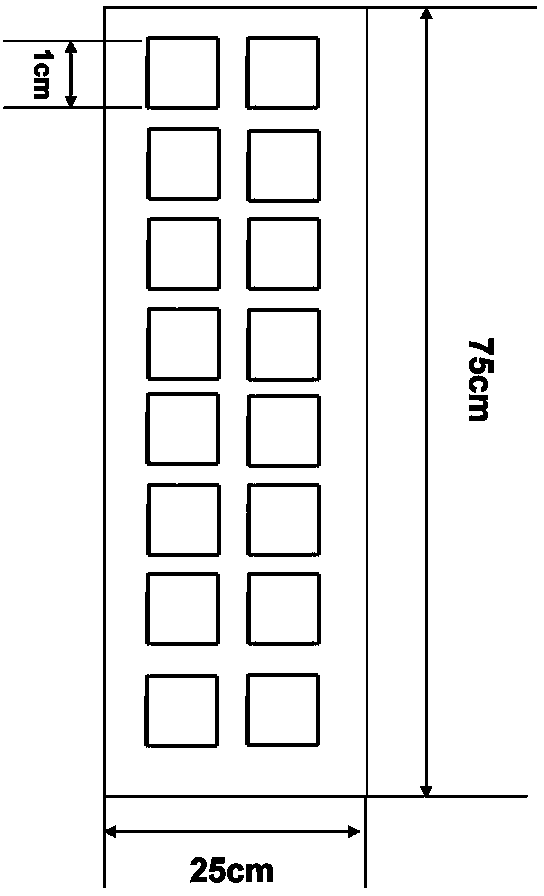
[0028] Step 2: Under the conditions of room temperature and relative humidity of 60%, use the NanoPlotter2.1 spotting instrument of German Gesim company to place 14 kinds of antigenic proteins (dsDNA, sm, P 0 ,P 1 ,P 2 , PCNA, U1-snRNP68 / 70, U-snRNP BB', U1-snRNP C, U1-snRNP A, Ro / SS-A 52KD, Ro / SS-A 60KD, La / SS-B, nucleolin) and 4 species Control substances (negative reaction control-antigen diluent, positive reaction control-goat anti-human IgG, positive display control-high concentration human IgG, negative display control-low concentration human IgG) were dotted on the cellulose acetate membrane according to the array arrangement. See the appendix for the spotting positions of each antigen protein and reference figure 1 , the dot spacing is 0.7mm, and the dot volume is about 10nL. Each sample i...
Embodiment 2
[0039] Detection of SLE autoantibodies:
[0040] Sample preparation: use the sample diluent in the systemic lupus erythematosus autoantibody detection device prepared in Example 1 to dilute the serum sample of a systemic lupus erythematosus patient at 1:500 to obtain a diluted sample;
[0041] Sample reaction: Take 1mL of the diluted sample, add it to the well of the multi-well plate, incubate with the chip, cover it, and shake it at room temperature for 1.5h;
[0042] Rinsing: Dilute the concentrated lotion 10 times with distilled water to obtain a rinsing solution, absorb the liquid in the wells of the plate, add 1 mL of the rinsing solution, shake for 5 minutes, absorb the liquid, and repeat the rinsing twice;
[0043] Enzyme-labeled secondary antibody binding: Dilute HRP-goat anti-human IgG at 1:1000 with the above washing solution, add 1 mL to each plate well, shake at room temperature for 1.5 h, absorb the liquid, then add 1 mL of washing solution, and keep at room tempe...
Embodiment 3
[0046] Collect 23 sera of SLE patients diagnosed with SLICC systemic lupus erythematosus standard in the Department of Rheumatology and Immunology of a tertiary hospital, and 30 sera of normal people in physical examination, apply the systemic lupus erythematosus autoantibody detection device of the present invention, refer to Example 2 The method detects. The test results showed that the detection rate of positive samples was 19 / 23=82.6%, and the detection coincidence rate of negative samples was 30-2 / 30=93.3% (both samples were positive for dsDNA). In the diagnostic criteria for systemic lupus erythematosus formulated by the International Society of Rheumatology, only 2 of the 11 reference indicators involve antibody abnormalities, and the diagnosis of the disease also needs to refer to multiple clinical symptoms of patients, so the detection device of the present invention can screen More than 80% of the disease patients were detected, indicating that the detection device h...
PUM
 Abstract
Description
Claims
Application Information
 - R&D
- Intellectual Property
- Life Sciences
- Materials
- Tech Scout
- Unparalleled Data Quality
- Higher Quality Content
- 60% Fewer Hallucinations
Browse by: Latest US Patents, China's latest patents, Technical Efficacy Thesaurus, Application Domain, Technology Topic, Popular Technical Reports.
© 2025 PatSnap. All rights reserved.Legal|Privacy policy|Modern Slavery Act Transparency Statement|Sitemap|About US| Contact US: help@patsnap.com


