Kit for quantitatively detecting CEA (Carcino-Embryonic Antigen) as well as preparation method and application method of kit
A quantitative detection and kit technology, applied in in vitro clinical testing and nanometer field, to achieve the effects of rapid response, high sensitivity and wide linear range
- Summary
- Abstract
- Description
- Claims
- Application Information
AI Technical Summary
Problems solved by technology
Method used
Image
Examples
Embodiment 1
[0030] Embodiment 1: Kit
[0031] This embodiment is a CEA quantitative detection kit, which is based on biofunctionalized magnetic nanocomposite particle technology, and has high sensitivity and rapid response. The so-called kit is a set product including multiple reagents, and the various reagents in the kit are used together to complete specific functions.
[0032] According to the present invention, the kit includes at least the following reagents: biofunctionalized magnetic nanocomposite particles coupled with CEA antibody, enzyme-labeled CEA antibody and chemiluminescent substrate solution. The biofunctionalized magnetic nanocomposite particles coupled with CEA antibody are used to capture CEA antigen and separate CEA from other media under the action of an external magnetic field; the enzyme-labeled CEA antibody is used to combine with CEA and biofunctionalized magnetic nanocomposite particles A sandwich structure is formed and the chemiluminescent substrate is catalyz...
Embodiment 2
[0040] Embodiment 2: the preparation method of kit
[0041] The preparation method of the kit includes the steps of preparing each reagent in the kit. In the present invention, it mainly lies in the preparation method of biologically functionalized magnetic nanocomposite particles, which is also the innovation and key of the present invention. Biologically functionalized magnetic nanocomposite particles are microspheres composed of superparamagnetic nanoparticles, and biological antibodies are coupled to the microspheres to form biofunctionalized magnetic nanocomposite particles.
[0042] In the first step, the biotinylated CEA antibody is added to the magnetic beads, and shaken for a period of time,
[0043]The two are coupled to form biofunctionalized magnetic nanocomposite particles; in the second step, BSA (bovine serum albumin) is added to the magnetic beads and the shaking reaction is performed again to block the unreacted sites on the magnetic beads; in the third step, ...
Embodiment 3
[0049] Embodiment 3: the method for using kit to detect CEA
[0050] 1. Pretreatment of kits and samples.
[0051] In this embodiment, the kit was placed at room temperature (18-26° C.) to equilibrate for 10-15 minutes; 100 μL of the concentrated washing solution was diluted with deionized water at 1:100 for later use; the samples to be tested (blood, pleural volume liquid, sputum, saliva, semen, urine, ascites), etc., take the supernatant, and put it at room temperature for 10-15 minutes to equilibrate.
[0052] 2. Using the kit to detect CEA on the sample.
[0053] Step 1: This step is the detection process of the calibrator, and the calibrator is used for calibration. This step consists of the following three steps:
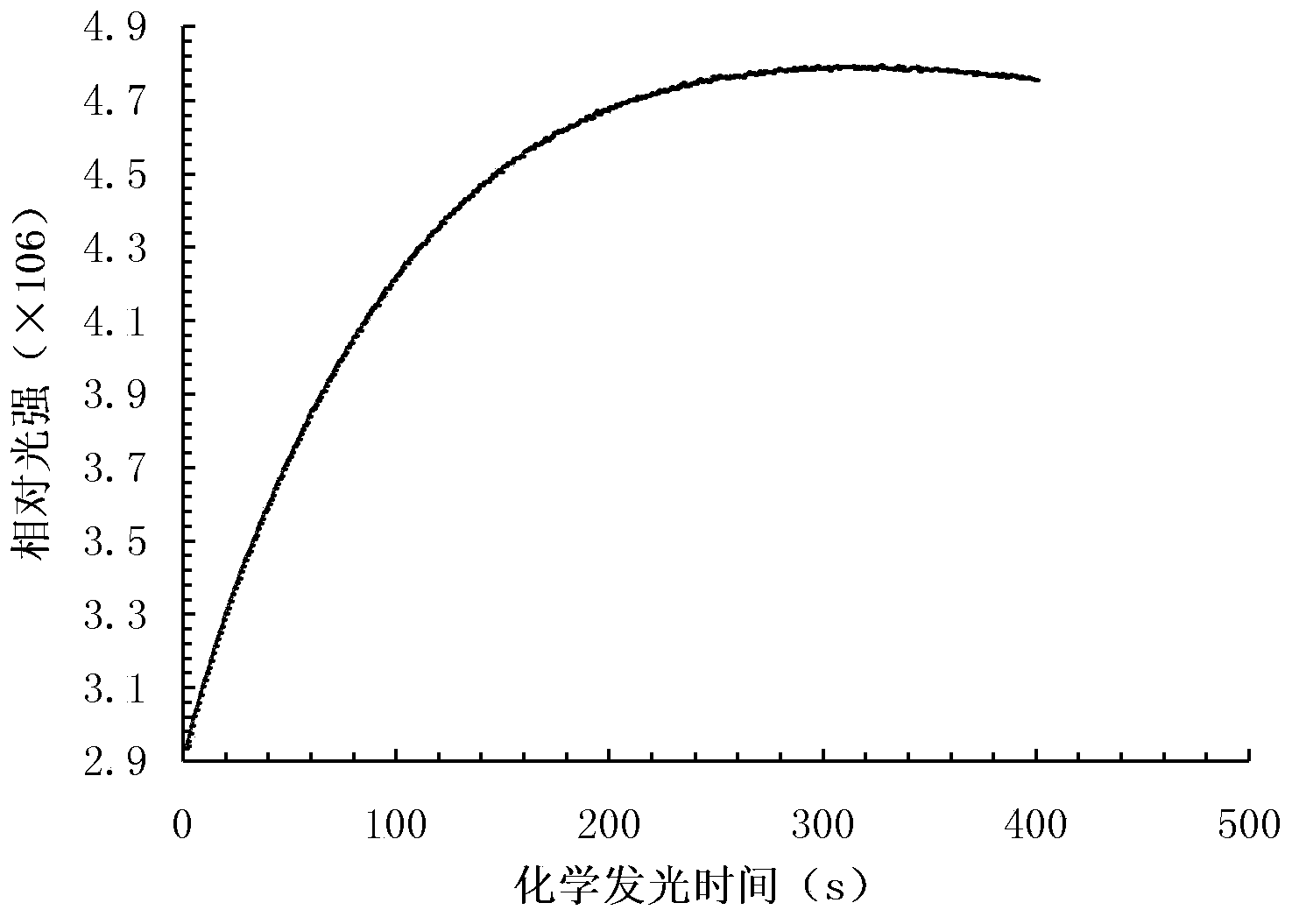
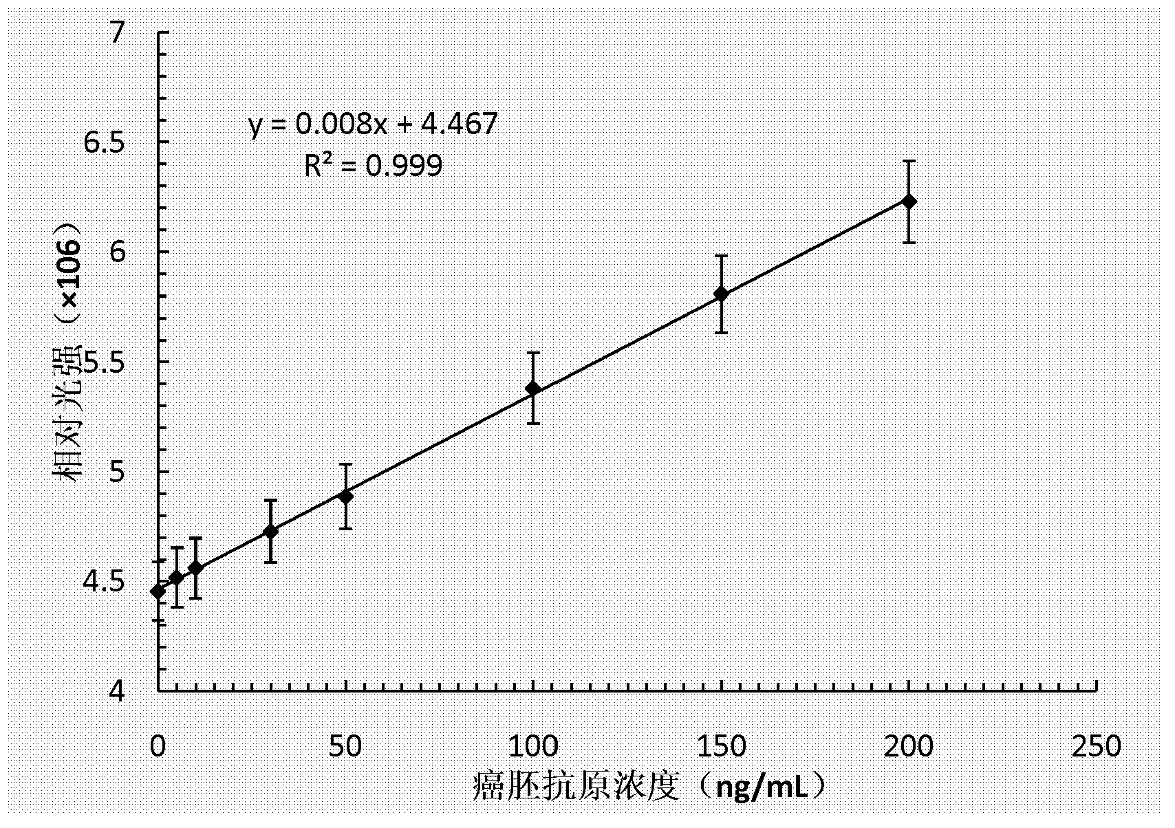
[0054] In the first step, the biofunctionalized magnetic nanocomposite particles are used in this step to capture the CEA in the solution. In this embodiment, 50 μL of biofunctionalized magnetic nanocomposite particles and 50 μL of calibrator are mixed, ta...
PUM
| Property | Measurement | Unit |
|---|---|---|
| concentration | aaaaa | aaaaa |
| molecular weight | aaaaa | aaaaa |
Abstract
Description
Claims
Application Information
 - R&D
- Intellectual Property
- Life Sciences
- Materials
- Tech Scout
- Unparalleled Data Quality
- Higher Quality Content
- 60% Fewer Hallucinations
Browse by: Latest US Patents, China's latest patents, Technical Efficacy Thesaurus, Application Domain, Technology Topic, Popular Technical Reports.
© 2025 PatSnap. All rights reserved.Legal|Privacy policy|Modern Slavery Act Transparency Statement|Sitemap|About US| Contact US: help@patsnap.com


