Method for evaluating biotoxicity of endosulfan
A technology of biotoxicity and endosulfan, applied in the biological field, can solve the problems of false positive, cell membrane damage, cell membrane structure damage, etc., and achieve the effect of simple method and high correlation
- Summary
- Abstract
- Description
- Claims
- Application Information
AI Technical Summary
Problems solved by technology
Method used
Image
Examples
Embodiment 1
[0034] The effect of embodiment 1 endosulfan on the cell proliferation of HUVEC-C
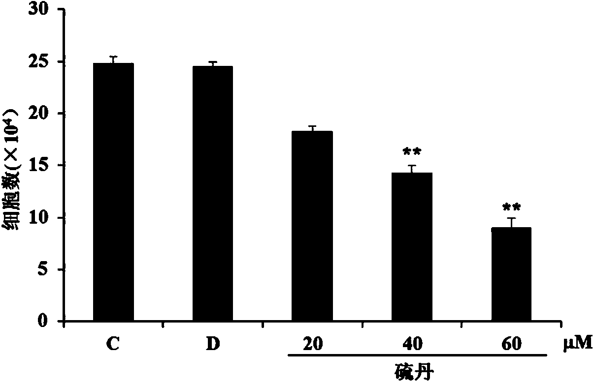
[0035] HUVEC-C (human umbilical vein endothelial cells) cells were exposed to endosulfan, and the experiment was divided into three groups: normal cell group (C), DMSO control group (dimethyl sulfoxide, D), endosulfan experimental group ( endosulfan, ES). Control group DMSO1% concentration, endosulfan exposure concentration is (20,40,60 μ M), exposure time is 48 hours, collects cells and carries out cell counting, as figure 1 The results showed that endosulfan exposure caused inhibition of cell proliferation in a dose-dependent manner. Compared with the control group, endosulfan significantly inhibited cell growth at a low concentration of 20 μM (P*<0.05), and significantly induced cell proliferation at a higher concentration (40-60 μM) Inhibition (P**<0.01).
Embodiment 2
[0036] The effect of embodiment 2 endosulfan on HUVEC-C cell cycle
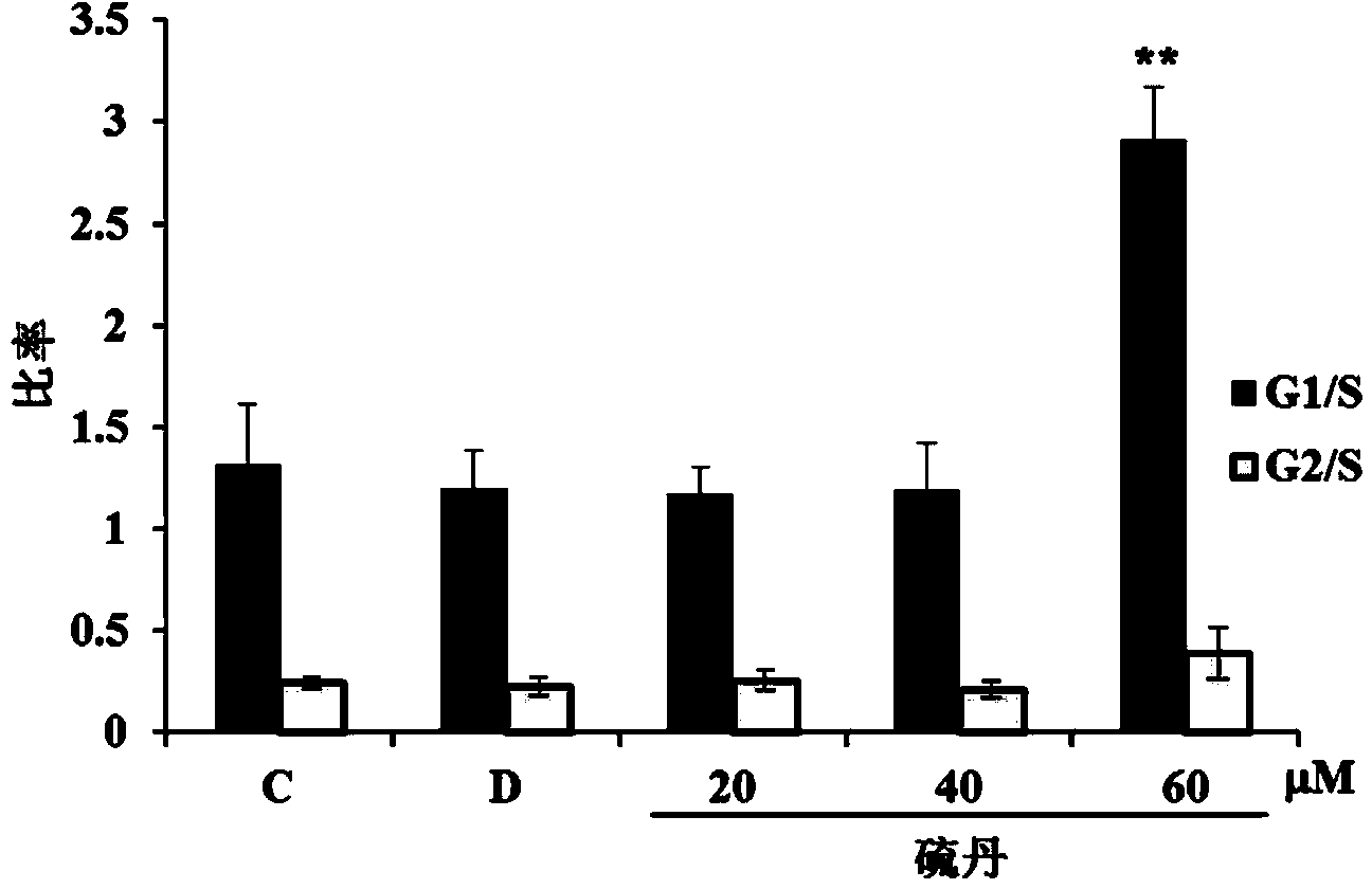
[0037] HUVEC-C cells were exposed to endosulfan for 48 hours, and the cells in the normal cell group, DMSO group, and endosulfan experimental group (20, 40, 60 μM) were collected, fixed with 70% cold ethanol overnight, and then stained with PI and RNase treatment, using flow cytometry (BD FACSCalibur) for cell cycle determination, such as figure 2 The results showed that when endosulfan was at a high concentration of 60 μM, compared with the control group, there was a G1 / S phase arrest, which was significant (P**<0.01).
Embodiment 3
[0038] Example 3 Exposure to endosulfan causes changes in the level of IL-6 secretion by cells
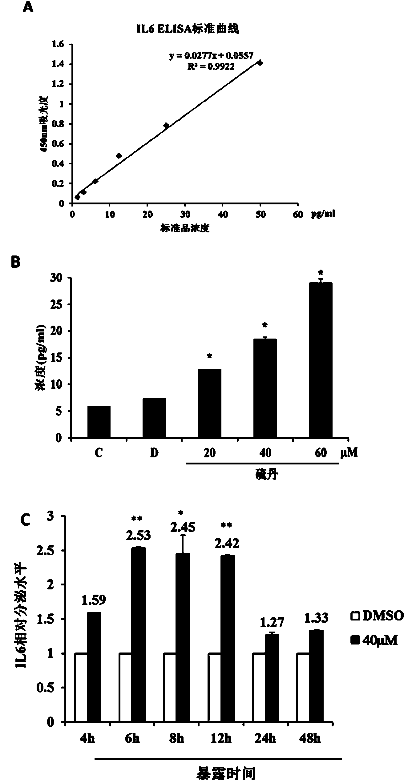
[0039] HUVEC-C cells have the characteristics of autocrine IL-6. First, in terms of dose effect, HUVEC-C cells were exposed to endosulfan for 12 hours, and the normal cell group, DMSO group, and endosulfan experimental group (20,40 , 60 μM) of the cell culture supernatants of the three groups were used to measure the level of IL-6 secreted by the cells using the IL-6human ELISA Kit (Abcam). image 3 A is the standard curve that utilizes ELISA method to measure IL-6, image 3 B is the effect of different concentrations of endosulfan on the secretion level of IL-6. image 3 The results of A and 3B show that exposure to endosulfan can increase the expression of IL-6, which is dose-dependent and statistically significant. Secondly, in terms of time effect, HUVEC-C cells were treated with different exposure times (4, 6, 8, 12, 24, 48 h), and the cell culture supernatants of the DMSO g...
PUM
 Abstract
Description
Claims
Application Information
 - R&D
- Intellectual Property
- Life Sciences
- Materials
- Tech Scout
- Unparalleled Data Quality
- Higher Quality Content
- 60% Fewer Hallucinations
Browse by: Latest US Patents, China's latest patents, Technical Efficacy Thesaurus, Application Domain, Technology Topic, Popular Technical Reports.
© 2025 PatSnap. All rights reserved.Legal|Privacy policy|Modern Slavery Act Transparency Statement|Sitemap|About US| Contact US: help@patsnap.com


