

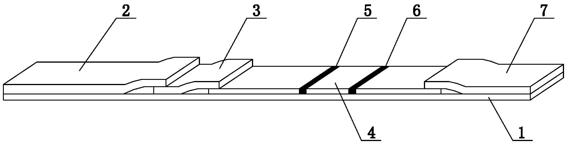
Test strip for rapidly detecting vitamin D
A vitamin and test strip technology, applied in biological testing, measuring devices, material inspection products, etc., can solve the problems of rare standard reference objects, unstable samples, complex vitamin D purification process, etc., to achieve convenient use and fill the gap in the market , low-cost testing effect
- Summary
- Abstract
- Description
- Claims
- Application Information
AI Technical Summary
Problems solved by technology
Method used
Image

Examples
Embodiment 1
[0036] Example 1 Preparation of conjugates of marker-labeled vitamin D (25-hydroxyvitamin D3) and carrier protein
[0037] Step 1. Dissolve 1 mg vitamin D (25OHD33-HS, purchased from Toronto Research Chemicals Company) in 100 μL organic solvent (n-hexane, acetonitrile, dimethyl sulfoxide or 1,4-dioxane), and then Add the dissolved vitamin D dropwise to 10 mL of carrier protein (bovine serum albumin BSA, or chicken ovalbumin OVA) solution with a concentration of 0.1 mg / mL, and then add pentadiene with a concentration of 50% by volume dropwise. Aldehyde aqueous solution until the volume percentage concentration of glutaraldehyde in the solution is 1%. After stirring for 5 minutes, move to 4°C and stir for 16 hours. The reaction solution after the reaction is dialyzed with PBS buffer solution for 8 hours at 4°C. ~3 times, after centrifuging the dialyzate, take the supernatant, and finally filter the supernatant with a 0.45 μm filter membrane to obtain a conjugate of vitamin D and...
Embodiment 2
[0039] Example 2 Preparation of conjugates of marker-labeled vitamin D (25-hydroxyvitamin D2) and carrier protein
[0040] Step 1. Dissolve 5 mg of vitamin D (25OHD2, purchased from Toronto Research Chemicals) in 100 μL of organic solvent (n-hexane, acetonitrile, dimethyl sulfoxide or 1,4-dioxane), and then Add the dissolved vitamin D dropwise to 10 mL of carrier protein (bovine serum albumin BSA, or chicken ovalbumin OVA) solution with a concentration of 2 mg / mL, and then add a 50% aqueous glutaraldehyde solution dropwise The volume percent concentration of glutaraldehyde in the solution is 2%. After stirring for 10 minutes, move to 4°C and stir for 17 hours. After the reaction, the reaction solution is dialyzed with PBS buffer solution at 4°C for 8 hours. During the dialysis, the liquid is changed for 2 to 3 The second time, the supernatant was taken after the dialyzate was centrifuged, and finally the supernatant was filtered with a 0.45 μm filter membrane to obtain a conju...
Embodiment 3
[0042] Example 3 Preparation of conjugates of marker-labeled vitamin D (25-hydroxyvitamin D3) and carrier protein
[0043] Step 1. Dissolve 10 mg of vitamin D (25OHD3, purchased from Toronto Research Chemicals) in 100 μL of organic solvent (n-hexane, acetonitrile, dimethyl sulfoxide or 1,4-dioxane), and then Add the dissolved vitamin D dropwise to 10 mL of carrier protein (bovine serum albumin BSA, or chicken ovalbumin OVA) solution with a concentration of 5 mg / mL, and then add a 50% aqueous glutaraldehyde solution drop by volume The volume percent concentration of glutaraldehyde in the solution is 3%. After stirring for 20 minutes, move to 4°C and stir for 18 hours. After the reaction, the reaction solution is dialyzed with PBS buffer solution at 4°C for 8 hours. The second time, the supernatant was taken after the dialyzate was centrifuged, and finally the supernatant was filtered with a 0.45 μm filter membrane to obtain a conjugate of vitamin D and carrier protein, which wa...
PUM
 Abstract
Description
Claims
Application Information