
Full-field optical coherence tomographic three-dimensional medical imaging device and method

A technology for optical coherence tomography and medical imaging, which is applied in the direction of material analysis, measurement device, and material analysis by optical means, which can solve the problems of reduced imaging quality, complex system, and difficult frequency, and achieve strong phase-shifting algorithm. The effect of adaptability, high system resolution, and reduced measurement time

Inactive Publication Date: 2015-04-29
NANJING UNIV OF SCI & TECH
View PDF2 Cites 21 Cited by
  • Summary
  • Abstract
  • Description
  • Claims
  • Application Information

AI Technical Summary

Problems solved by technology

The acquisition speed of this method is fast, but the disadvantage is that the system is complex and costly, and the biological tissue is almost transparent under near-infrared light illumination, which will result in a decrease in imaging quality
Another way is to generate sinusoidal signals through piezoelectric ceramics and trigger CCD externally. Using the characteristics of high-speed exposure of CCD, a set of acquisitions can be calculated to obtain the enface map. This method relies on the signal generated by piezoelectric ceramics to match the CCD. In practice, It is difficult to ensure simultaneous operation during operation and the frequency cannot be changed as needed
[0005] The current intraoperative sudden pathological diagnosis uses frozen sections to process tissues, and then stain and label them. Not only will these chemical treatments pollute the tissues, but the time required for the preparation process will also cause patients to miss the best treatment period

Method used


Image

  • Full-field optical coherence tomographic three-dimensional medical imaging device and method
  • Full-field optical coherence tomographic three-dimensional medical imaging device and method
  • Full-field optical coherence tomographic three-dimensional medical imaging device and method

Examples


Embodiment Construction

[0020] The present invention will be described in further detail below in conjunction with the accompanying drawings and specific embodiments.

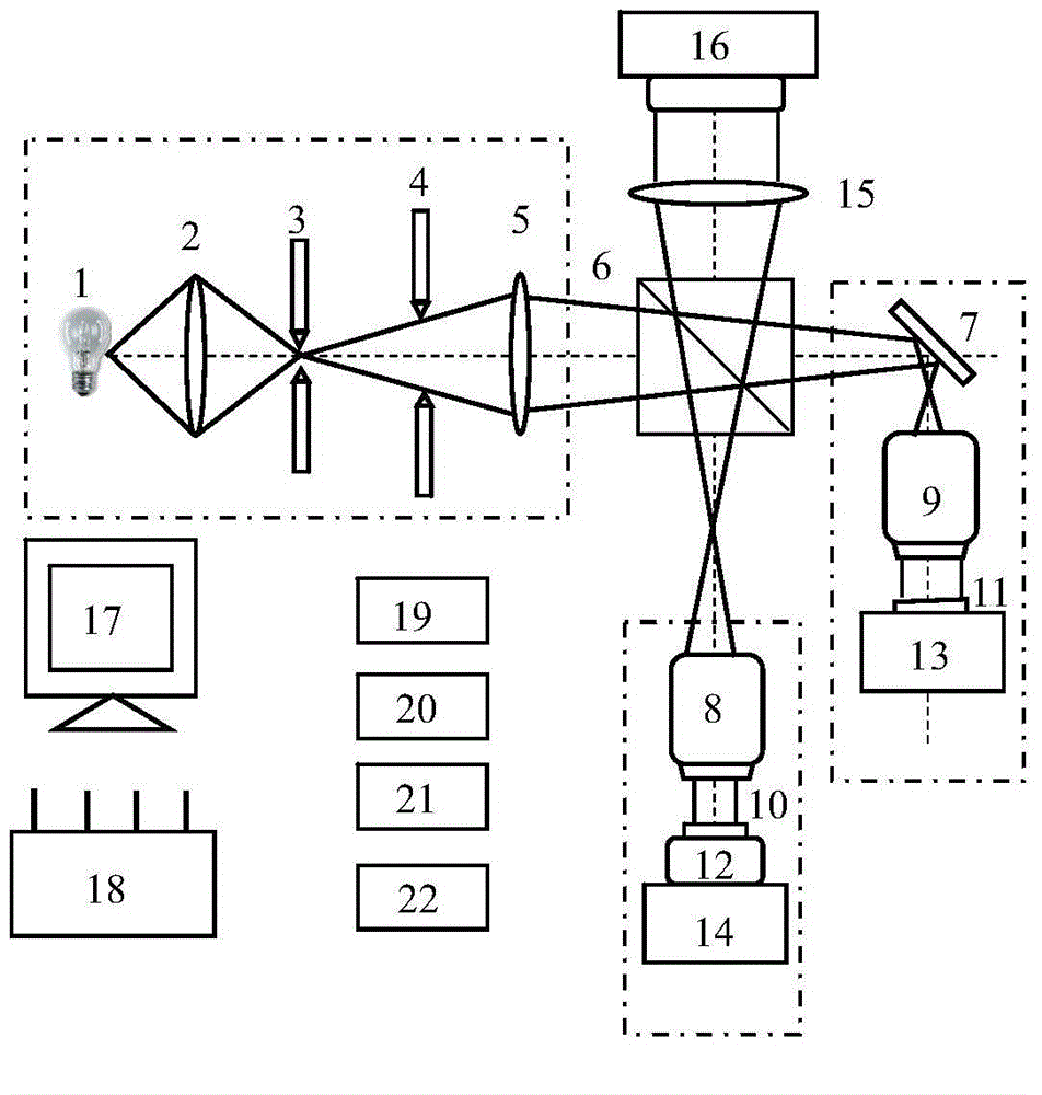
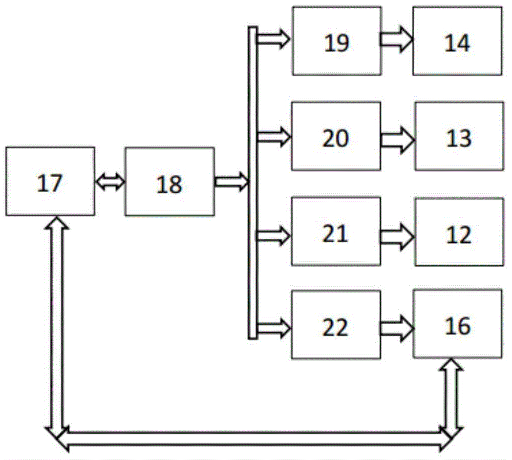
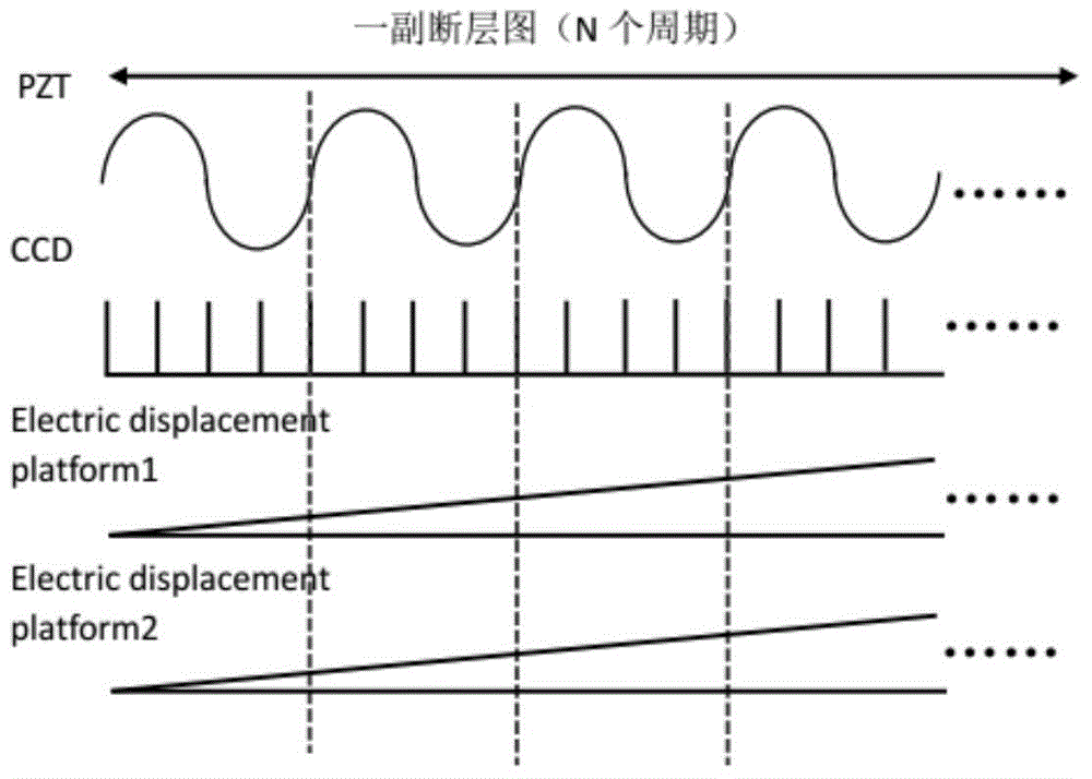
[0021] combine Figure 1~2 , the full-field optical coherence tomography three-dimensional medical imaging device of the present invention includes a tungsten-halogen light source 1, a front condenser lens 2, an aperture stop 3, a field stop 4, a first converging lens 5, a dichroic prism 6, and a reflector 7 , reference arm microscope objective lens 8, sample arm microscope objective lens 9, reference mirror 10, sample 11, piezoelectric ceramics 12, sample arm electronically controlled displacement platform 13, reference arm electrically controlled displacement platform 14, second converging lens 15, surface Array CCD16, computer 17, single-chip microcomputer 18, reference arm electric displacement platform controller 19, sample arm electric displacement platform controller 20, piezoelectric ceramic controller 21, CCD controller 22; s...


PUM

PropertyMeasurementUnit
Center wavelengthaaaaaaaaaa
Bandwidthaaaaaaaaaa

Abstract

The invention discloses a full-field optical coherence tomographic three-dimensional medical imaging device and a full-field optical coherence tomographic three-dimensional medical imaging method. The device comprises a Kohler illumination part, a reference arm, a sample arm, an imaging lens and an area array CCD detector; according to the method, piezoelectric ceramics are adopted for phase shift of a reference mirror according to the white light low-coherence interference theory; the optical path of the two arms is changed; CCD is used for receiving an interference signal; the interference signal is processed by a computer to obtain a two-dimensional fault map of a sample; finally an electric control displacement platform is used for axial scanning to obtain three-dimensional information, wherein a single chip can be used for emitting two different signal modulation piezoelectric ceramics; the CCD and the electric control displacement platform for the two arms correspond to two demodulation modes. According to the device and the method disclosed by the invention, the sample needs not to be subjected to the treatments such as slicing, dyeing and fluorescence labeling; frozen sections and paraffin sections which have the defects such as complex preparation and low resolution ratio can be replaced; a handhold medical instrument can be made for submicron-grade in-vitro imaging or in-vivo imaging of biological tissues.

Description

technical field [0001] The invention relates to the technical field of a full-field optical coherence tomography three-dimensional medical imaging system (Full-field optical coherence tomography, FF-OCT), in particular to a full-field optical coherence tomography three-dimensional medical imaging device and method. Background technique [0002] Full-field optical coherence tomography (Full-field Optical Coherence Tomography, FF-OCT) was first proposed in 1998. It is a non-invasive imaging technology that can obtain micron-scale in vivo optical tomographic imaging of biological tissues. It has important application prospects in the field of biomedicine. The FF-OCT system is famous for its fast parallel scanning mode. One scan can obtain the two-dimensional information of the X-Y plane, and the three-dimensional structure can be obtained by scanning in the depth direction of the sample arm. Combined with a large NA microscope objective lens and a broadband light source, FF-OC...

Claims


Application Information

Patent Timeline
IPC IPC(8): G01N21/84A61B5/00
Inventor 高万荣朱越廖九零陈朝良卞海溢伍秀玭刘浩
Owner NANJING UNIV OF SCI & TECH
Learn More