Method for detecting embryo chromosome abnormalities by using blastula-stage embryo cells

A technology for chromosomal abnormalities and embryonic cells, which is applied in the field of chromosomal abnormality detection, can solve the problems of affecting experimental results, small number of templates, cumbersome operations, etc., and achieve the effect of improving amplification effect, increasing success rate, and high detection throughput

Inactive Publication Date: 2015-06-17
SUZHOU BASECARE MEDICAL DEVICE CO LTD
View PDF4 Cites 27 Cited by
  • Summary
  • Abstract
  • Description
  • Claims
  • Application Information

AI Technical Summary

Problems solved by technology

At present, clinical PGS detection methods mainly include Fish, Array CGH, etc. However, these two technologies have certain limitations: the current problem of FISH technology is that it cannot detect all chromosomes at one time, the operation is cumbersome, and the resolution is low; CGH The disadvantage is that it can only detect known chromosomal abnormalities and is expensive
No matter what kind of detection technology is based on, it is first necessary to separate part of the cells from the developing embryo in vitro. At present, the blastomere separation method is commonly used to separate 1-2 blastomeres from embryos that have developed to the 8-cell stage. Chromosomal abnormalities are detected by single-cell amplification sequencing of 1-2 blastomeres, but due to the extremely small number of templates, amplification failures and large amplification biases are often prone to problems, which affect the experimental results

Method used


Image

  • Method for detecting embryo chromosome abnormalities by using blastula-stage embryo cells
  • Method for detecting embryo chromosome abnormalities by using blastula-stage embryo cells
  • Method for detecting embryo chromosome abnormalities by using blastula-stage embryo cells

Examples


Embodiment 1

[0020] Isolation of blastocyst trophectoderm cells

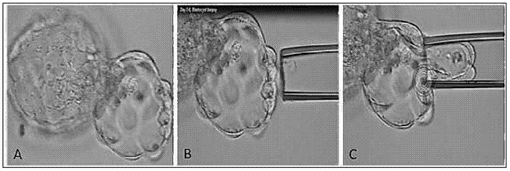
[0021] Select in vitro fertilized embryos from high-risk groups (age ≥ 35, repeated implantation failure, repeated miscarriage) in IVF, and obtain blastocyst trophectoderm cells by conventional embryo biopsy methods; when the embryo develops to the D3 stage, the zona pellucida is used to infiltrate Well, continue to culture until D5 stage, when trophectoderm cells extrude the zona pellucida, such as figure 1 , absorb part of the trophectoderm cells (5-10) with a flat micromanipulator needle, and cut off with a laser; the absorbed cells are transferred to PBS solution and washed 3 times.

Embodiment 2

[0023] Cell Lysis and Single Cell Expansion

[0024] (1) Cell Lysis

[0025] Transfer the washed cells to a 0.2mL PCR tube. According to the number of samples, mix the cell lysis buffer and cell lysis enzyme, and add the mixture to the cell samples. The reaction system for a single sample is shown in Table 1 below:

[0026] Table 1

[0027] components

Reaction volume (μL)

single cell sample

<2

Cell Lysis Buffer

5

Cytolytic enzyme

0.1

Total amount of reaction system

<7.1

[0028] Incubate the samples in a preheated PCR machine under the conditions shown in Table 2 below:

[0029] Table 2

[0030]

[0031] (2) Pre-amplification

[0032] According to the number of samples, mix the pre-amplification buffer and pre-amplification enzyme, and add the mixture to the reaction tube in the previous step. The single-sample reaction system is shown in Table 3 below:

[0033] table 3

[0034] components

Reac...

Embodiment 3

[0048] DNA fragmentation

[0049](1) DNA Fragmentation Use NEB's Fragmentation Enzyme Kit, briefly centrifuge the DNA fragmentation enzyme for 3 seconds, place it on an ice box, and configure the system shown in Table 7 below in a 1.5mL EP tube:

[0050] Table 7

[0051] components

Reaction volume (μL)

Purified DNA samples

X (500ng)

fragmentation buffer

2

water

16-X

Fragmentase

2

Total amount of reaction system

20

[0052] Place the EP tube in a thermostatic metal bath at 37°C for 30 min; after the reaction, add 5 μL of stop reaction buffer.

[0053] (2) DNA purification after fragmentation

[0054] Add 30 μL of water to the reaction system to make the volume of the system reach 50 μL, add 75 μL of XP magnetic beads, shake the Votex or blow it with a gun to mix the reaction system, and let it stand at room temperature for 5 minutes; put it into the magnetic stand, wait until the magnetic beads are co...


PUM


Abstract

The invention discloses a method for performing genome amplification by using embryo blastula-stage cells, performing chromosome detection on the preimplantational embryo by combining a high-flux sequencing technique and screening out the chromosome normal embryo. The method can comprehensively and completely analyze the genetic variation information of the embryo genome, thereby instructing the preimplantational embryo selection, reducing the hereditary diseases and enhancing the success rate of test tube babies. The method comprises the following steps: blastula-stage trophocyte separation; genome amplification; DNA (deoxyribonucleic acid) segmentation; and Proton library establishment, mounting sequencing and sequencing data analysis. By using the blastula-stage embryo to perform trophocyte separation detection, the method avoids the injuries of cleavage-stage cell separation to the embryo, obtains higher cell quantity than the cleavage stage, and enhances the success rate and amplification effect of genome amplification. After the blastula-stage embryos are subjected to the natural elimination process, the high-quality blastula-stage embryo is selected for detection, thereby saving the cost.

Description

technical field [0001] The invention relates to the field of biomedicine, in particular to a method for detecting chromosomal abnormality on the basis of single cell amplification using embryonic cells developed to the blastocyst stage in vitro. Background technique [0002] Twenty years ago, the infertility rate among people of childbearing age in my country was only 3%, which was at a relatively low level in the world. The 2009 China Infertility Summit Forum released the "China Infertility Status Survey Report", which shows that the number of infertility patients in the country has exceeded 50 million, and the number of infertility patients aged 25 to 30 is the largest, showing a younger trend. One out of eight couples of childbearing age faces difficulties in childbearing, and the infertility rate climbs to 12.5%-15%, which is close to the rate of 15%-20% in developed countries. The most serious thing is that this ratio is still rising. Experts from the World Health Orga...

Claims


Application Information

Patent Timeline
Patent Type & Authority Applications(China)
IPC IPC(8): C12Q1/68
Inventor 梁波孔令印冒燕宣黎明申静静祝轶君
Owner SUZHOU BASECARE MEDICAL DEVICE CO LTD
Why Eureka
Try Eureka