A poisonous microcarbon algae crack algae and its separation method and application
A technology of cyanophage and Microcystis, which is applied in the field of pyrolysis and separation of toxic Microcystis cyanophage, can solve the problems of consuming a lot of manpower and material resources, and chemical agent pollution, and achieve rapid lysis, strong specificity, and high cultivation conditions simple effect
- Summary
- Abstract
- Description
- Claims
- Application Information
AI Technical Summary
Problems solved by technology
Method used
Image
Examples
Embodiment 1
[0026] Preparation of cyanophage MaSSC-P
[0027] The water sample was collected from Ningbo Bang Cultural Park, Ningbo City, Zhejiang Province; the host algae was Microcystis aeruginosa M. aeruginosa FACHB-925, the specific preparation method is as follows:
[0028] (1) Isolation of cyanophages
[0029] The collected surface water samples containing toxic Microcystis lysed cyanophages were filtered through medium-speed filter paper, 0.45 μm and 0.22 μm nitrocellulose filter membranes; the filtrate was mixed with Microcystis aeruginosa in logarithmic growth phase by volume Infection mixture at a ratio of 1:4; at the same time, the water sample (without cyanophage particles) filtered through a tangential flow 10KD membrane was infected with Microcystis aeruginosa in the logarithmic growth phase at the same ratio as a control; 2000 lx, the temperature is 25 ℃, and the light-dark cycle is 12h:12h; the yellowed culture solution is continuously subcultured until the cyanophage...
Embodiment 2
[0038] Morphological Observation of Lysing Cycphages of Microcystis aeruginosa
[0039] Take the cyanophage isolated and purified in Example 1 on the copper grid, negatively stain with 2% uranyl acetate solution (w / v), and then observe the cyanophage under an electron microscope (Hitachi H-7650). form.
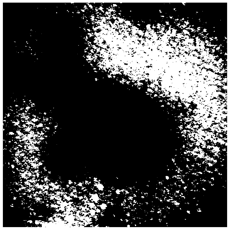
[0040] The result is as figure 1 As shown, the cyanophage has a regular polyhedral head structure and a slender tail, the diameter of the head is about 85nm, and the length of the tail is about 250nm.
[0041] The purified cyanophages were infected and mixed with exponentially growing Microcystis aeruginosa, and the algal plaque experiment was carried out, and the algal plaques with uniform size and shape could be obtained, without halos around, and clear and regular edges.
Embodiment 3
[0043] Nucleic acid type identification of cyanophage genome
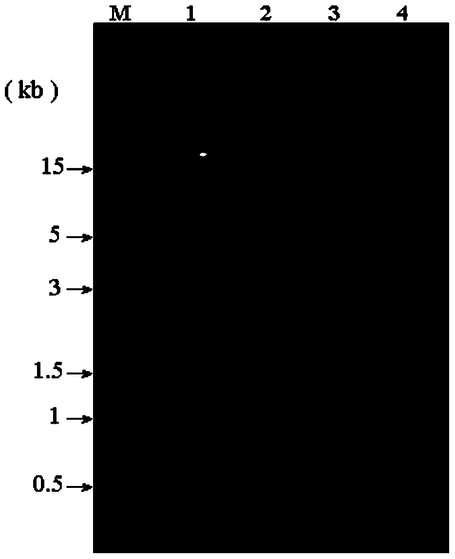
[0044] Take the cyanophage particles separated and purified in Example 1, and after extracting the nucleic acid, use DNase I, Rnase A and S1 single-stranded nuclease to act on the cyanophage genome nucleic acid respectively. The action spectrum is as follows figure 2 As shown in the figure (Swimming lane 1: Untreated nucleic acid, Swimming lane 2: Dnase I enzyme treatment, Swimming lane 3: Rnase A enzyme treatment, Swimming lane 4: S1 single-strand enzyme treatment) the cyanophage can be detected by Dnase I, single-strand Nuclease digestion, but cannot be digested by RNase A enzyme, and then it is concluded that cyanophage MaSSC-P is a virus particle containing single-stranded DNA (ssDNA); according to the "Virus Classification- According to the Eighth Report of the International Committee on Taxonomy of Viruses, the cyanophage does not belong to Myoviridae, Longoviridae and Brachyviridae, and should represent a n...
PUM
| Property | Measurement | Unit |
|---|---|---|
| diameter | aaaaa | aaaaa |
Abstract
Description
Claims
Application Information
 - R&D
- Intellectual Property
- Life Sciences
- Materials
- Tech Scout
- Unparalleled Data Quality
- Higher Quality Content
- 60% Fewer Hallucinations
Browse by: Latest US Patents, China's latest patents, Technical Efficacy Thesaurus, Application Domain, Technology Topic, Popular Technical Reports.
© 2025 PatSnap. All rights reserved.Legal|Privacy policy|Modern Slavery Act Transparency Statement|Sitemap|About US| Contact US: help@patsnap.com


