Spectral pupil laser differential confocal LIBS, Raman spectrum-mass spectrum microscopic imaging method and Raman spectrum-mass spectrum microscopic imaging device

A technique of differential confocal and Raman spectroscopy, applied in the directions of Raman/scattering spectroscopy, spectrometry/spectrophotometry/monochromator, microscope, etc. Problems such as low spatial resolution and drift in group detection and mass spectrometry detection can achieve the effects of overcoming stray light interference at the focal plane, improving spatial resolution, and strong anti-stray light ability

Inactive Publication Date: 2016-01-13
BEIJING INSTITUTE OF TECHNOLOGYGY
View PDF5 Cites 24 Cited by
  • Summary
  • Abstract
  • Description
  • Claims
  • Application Information

AI Technical Summary

Problems solved by technology

[0005] 1) Due to the use of simple laser focusing to desorb and ionize the sample, there are still problems such as large laser focusing spot and low spatial resolution of mass spectrometry detection;
[0006] 2) It is impossible to detect neutral atoms, molecules, neutral ions and groups, etc., which restricts the accurate and complete acquisition of sample component information;
[0007] 3) The time required for mass spectrometry imaging is long, and the axial position of the focused spot of the laser mass spectrometer often drifts relative to the sample to be measured

Method used


Image

  • Spectral pupil laser differential confocal LIBS, Raman spectrum-mass spectrum microscopic imaging method and Raman spectrum-mass spectrum microscopic imaging device
  • Spectral pupil laser differential confocal LIBS, Raman spectrum-mass spectrum microscopic imaging method and Raman spectrum-mass spectrum microscopic imaging device
  • Spectral pupil laser differential confocal LIBS, Raman spectrum-mass spectrum microscopic imaging method and Raman spectrum-mass spectrum microscopic imaging device

Examples


Embodiment 1

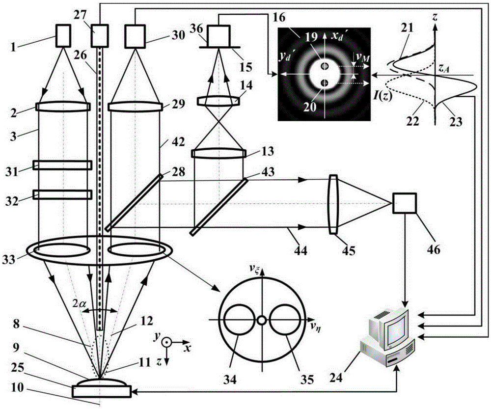
[0049] Such as figure 2 In the shown split-pupil laser differential confocal LIBS and Raman spectrum-mass spectrometer imaging device, the compressed focusing spot system 4 is replaced by a vector beam generation system 31 and a pupil filter 32, and the D-type illumination collector mirror 5 can be replaced by a circular Shaped illumination collecting mirror 33 is replaced, and first intensity point detector 17 and second intensity point detector 18 are replaced by CCD detector 36.

[0050] Such as figure 2 The shown split-pupil laser differential confocal LIBS and Raman spectroscopy-mass spectroscopy microscopic imaging device includes a point light source 1, a collimator lens 2 placed along the direction of the incident optical axis 8, a vector beam generating system 31, and a pupil filter 32 and focus spot to the circular illumination pupil 34 of the circular illumination collection mirror 33 of the tested sample 9, also includes the circular collection pupil 35 of the c...

Embodiment 2

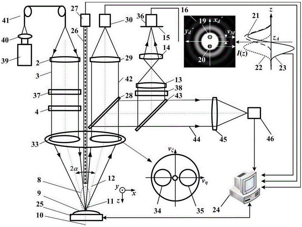
[0072] Such as image 3 In the shown split-pupil laser differential confocal LIBS and Raman spectrum-mass spectrometer imaging device, the point light source 1 is replaced by a pulsed laser 39, a condenser lens 40, and a light-transmitting optical fiber 41 at the focal point of the condenser lens 40, D The type illumination collecting mirror 5 is replaced by a circular illuminating collecting mirror 33 , and the first intensity point detector 17 and the second intensity point detector 18 are replaced by a CCD detector 36 . At the same time, an exit beam attenuator 37 is introduced into the laser focusing system, and a detection beam attenuator 38 is introduced into the laser split pupil differential confocal detection system.

[0073] The light intensity adjustment system is composed of the outgoing beam attenuator 37 and the detection beam attenuator 38, which are used to attenuate the focused spot and the spot intensity detected by the CCD detector 36, so as to meet the ligh...


PUM


Abstract

The invention relates to a spectral pupil laser differential confocal LIBS, a Raman spectrum-mass spectrum microscopic imaging method and a Raman spectrum-mass spectrum microscopic imaging device and belongs to the technical fields of confocal microscopic imaging, optical-spectrum imaging and mass spectrum imaging. In the invention, spectral pupil laser differential confocal imaging is combined with optical-spectrum and mass spectrum detection technologies, so that high-spatial-discrimination form imaging to a sample is carried out by means of a micro focus light spot of a spectral pupil laser differential confocal microscope which is subjected to ultra-discrimination technology treatment; mass spectrum detection to charged molecules and atoms in a micro zone of a sample is carried out by means of the mass spectrum detection system; micro zone optical spectrum detection is carried out to a focused light spot excitation spectrum (Raman spectrum, induced breakdown spectrum) of the spectral pupil laser differential confocal microscopic system through the optical-spectrum detection system; and high-spatial discrimination, high-sensitive imaging and high-sensitive detection to complete component information and form parameters of the sample micro zone is carried out through advantage complement and structural fusion of laser multi-spectrum detection. The invention provides a novel technical approach for imaging detection of substance components and formations in the field of biology, material and the like.

Description

technical field [0001] The invention belongs to the field of confocal microscopic imaging technology, spectral imaging technology and mass spectrometric imaging technology, and combines the split-pupil differential confocal microscopic imaging technology, laser-induced breakdown spectral imaging technology, Raman spectral imaging technology and mass spectroscopic imaging technology, and relates to a Split-pupil laser differential confocal LIBS, Raman spectroscopy-mass spectroscopy microscopic imaging methods and devices have broad application prospects in the fields of biology, materials, minerals, and micro-nano manufacturing. technical background [0002] Mass spectrometry (Mass Spectrometry) is to ionize the components in the sample, so that the generated charged atoms, molecules or molecular fragments with different charge-to-mass ratios are respectively focused under the action of electric field and magnetic field to obtain a spectrum arranged in order of mass-to-charge ...

Claims


Application Information

Patent Timeline
Patent Type & Authority Applications(China)
IPC IPC(8): G01N21/63G01N21/65G01N27/64
CPCG01J3/44G01N21/65G01N21/718G02B21/0032G02B21/0052G02B21/0076G02B21/008G02B21/20H01J49/0004G01J3/4412G01J3/443
Inventor 赵维谦王允邱丽荣
Owner BEIJING INSTITUTE OF TECHNOLOGYGY
Why Eureka
Try Eureka