Microlens imaging-based antigen antibody reaction determination method

A technology of antigen-antibody and determination method, applied in the direction of phase influence characteristic measurement, etc.

Active Publication Date: 2016-02-17
JINAN UNIVERSITY
View PDF5 Cites 3 Cited by
  • Summary
  • Abstract
  • Description
  • Claims
  • Application Information

AI Technical Summary

Problems solved by technology

However, the main technologies currently used in immunoassays (such as ELISA assay technology, SPR technology, fluorescent immunoassay, c

Method used


Image

  • Microlens imaging-based antigen antibody reaction determination method
  • Microlens imaging-based antigen antibody reaction determination method
  • Microlens imaging-based antigen antibody reaction determination method

Examples


Embodiment 1

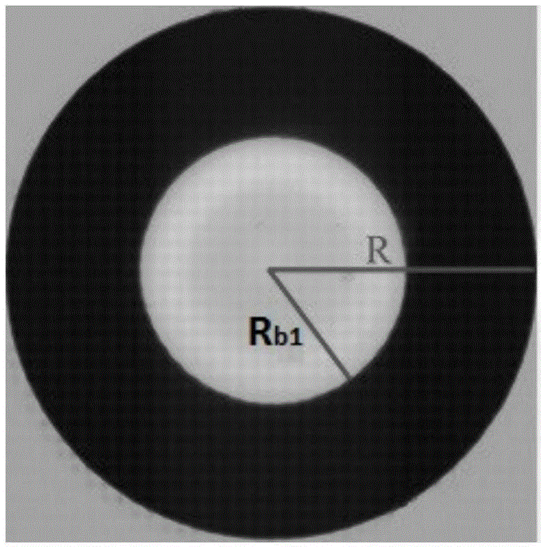
[0047] In this embodiment, one end is a spherical surface, and the other end is a flat microlens imaging technology to measure the reaction between ODA antibody and ODA serum: a silicon dioxide microlens is placed in a hole of a multi-well plate, and then ODA serum is dropped into the microlens. Immersion, at this time, use a digital imaging system to image the microlens illuminated by parallel light, and use special image analysis software to measure the radius R of the microlens and the radius R of the bright spot in the image center b . Then drop in the antibody solution, take a picture of the microlens every two seconds immediately after the drop, obtain 50 images of the microlens within 100 seconds, and measure the radius R of the central bright spot in each image b , together with the radius R of the microlens, use Equation (2) to calculate the refractive index of the solution, take the average of the refractive index values ​​from the 10th second to the 50th second, and...

Embodiment 2

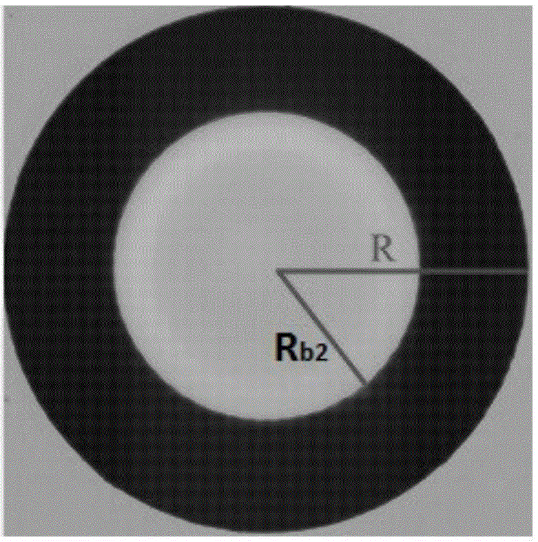
[0049] In this example, the reaction between CRP antibody and CRP antigen is measured by microsphere microlens imaging technology: a polystyrene microlens is placed in a porous plate, and then the CRP antigen solution is dropped to immerse the microlens. At this time, digital imaging is used to The system imaging images the microlens illuminated by parallel light, and uses special image analysis software to measure the radius R of the microlens and the radius R of the bright spot in the image center b . Then drop in the antibody solution, take a picture of the microlens every second immediately after dropping in, obtain 100 images of the microlens within 100 seconds, and measure the radius R of the central bright spot in each image b , together with the radius R of the microlens, use equation (1) to calculate the refractive index of the solution, take the average of the refractive index values ​​from the 10th second to the 80th second, and then quantitatively determine the ant...

Embodiment 3

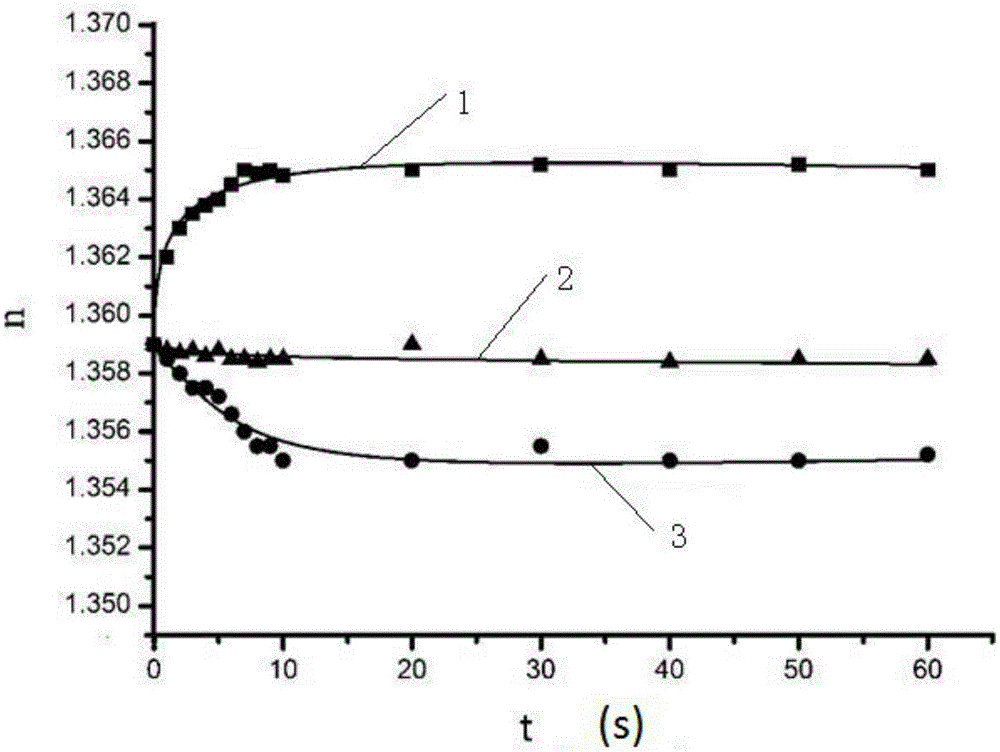
[0051] In this embodiment, one end is a spherical surface, and the other end is a flat microlens imaging technology to measure the reaction of interferon antibody and its antigen: a polystyrene microlens is placed in a porous plate, and then the interferon antigen solution is dropped into the microlens. Immersion, at this time, use the digital imaging system to image the microlens illuminated by parallel light, and use special image analysis software to measure the radius R of the microlens and the radius R of the bright spot in the image center b . Then drop in the antibody solution, take a picture of the microlens every second immediately after dropping in, obtain 40 images of the microlens within 600 seconds, and measure the radius R of the central bright spot in each image b , together with the radius R of the microlens, the curve of the solution refractive index changing with the antigen-antibody reaction process (such as Figure 4 shown), and then use the reaction balan...


PUM


Abstract

The invention discloses a microlens imaging-based antigen antibody reaction determination method. According to the microlens imaging-based antigen antibody reaction determination method, a microlens is immersed in a sample solution containing antigens, and under parallel light irradiation, an image is obtained, wherein the obtained image is a bright circle with a dark peripheral edge ring; an antibody solution is added dropwise, solution refractive index is changed after antigen antibody reaction, so that the thickness of the microlens image dark ring is changed; the thickness changing is determined, a formula derived from geometric optical principles is used for calculating the change of the solution refractive index after antigen antibody reaction, a curve used for representing the relationship of the changing amount with the concentration of the antigen is constructed, and it can be qualitatively determined whether antigen antibody reaction is induced, and antigen concentration and reaction dynamic parameters can be obtained via quantitative determination. Operation of the microlens imaging-based antigen antibody reaction determination method is simple and rapid; the microlens imaging-based antigen antibody reaction determination method can be used for realizing qualitative and quantitative determination of antigen antibody reaction; coating of a solid carrier with the antigen or the antibody before detection is not needed; and washing after detection is not needed.

Description

technical field [0001] The invention relates to the technical field of antigen-antibody reaction measurement, in particular to an antigen-antibody reaction measurement method based on microlens imaging. Background technique [0002] Antigen-antibody reaction detection and analysis is one of the most important detection and analysis methods in biology, basic medical research and clinical research, as well as food safety and environmental monitoring. At present, the most important technologies for antigen-antibody reaction detection and analysis include enzyme-linked immunosorbent assay (ELISA), surface plasmon resonance (SPR) technology, radioimmunoassay (RIA) method, fluorescence immunoassay and chemiluminescence immunoassay, etc. ELISA assay technology combines the amplification effect of enzyme-catalyzed reaction with the high specificity and specificity of antigen-antibody affinity reaction, and uses enzyme-labeled antigen or antibody as the main reagent for immunoassay, ...

Claims


Application Information

Patent Timeline
IPC IPC(8): G01N21/41
CPCG01N21/41
Inventor 黄耀熊
Owner JINAN UNIVERSITY
Why Eureka
Try Eureka