Monoclonal antibody against porcine epidemic diarrhea virus N protein and application thereof
A porcine epidemic diarrhea and monoclonal antibody technology, which is applied in antiviral immunoglobulins, antiviral agents, antibodies, etc., can solve the problems of labor consumption, inability to be widely used, time-consuming diagnostic techniques, etc., and achieve good application prospects and good reactive effects
- Summary
- Abstract
- Description
- Claims
- Application Information
AI Technical Summary
Problems solved by technology
Method used
Image

Examples
Embodiment 1
[0035] Example 1 Recombinant expression of porcine epidemic diarrhea virus N protein
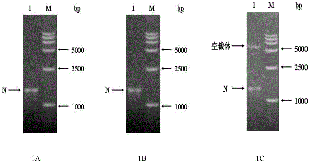
[0036] According to the nucleotide sequence information of porcine epidemic diarrhea virus JS-2013 strain, a pair of primers for amplifying the full length of the N gene (1326bp, the full length sequence of the N gene is shown in SEQ ID NO.2) was designed, provided by Shanghai Sangon Bioengineering Co., Ltd. Ltd Synthetics. The sequence of the upstream primer is: N-F: 5'-GCTCGGTACCCTCGAGATGGCTTCTGTCAGTTTTCAGGATC-3' (SEQ ID NO.3); the sequence of the downstream primer is N-R: 5'-ATTCGGATCCCTCGAGTTAATTTCCTGTGTCGAAGATCTCG-3' (SEQ ID NO.4), and the restriction site is XhoI. Extract viral genome RNA, reverse transcribe and synthesize cDNA, use primers N-F, N-R to amplify the full-length N gene, detect and identify the PCR product by 1% agarose gel electrophoresis, and then amplify the N gene with a size of about 1300bp by PCR. The expected result matches ( figure 1 A). Carrier pColdIDNA was di...
Embodiment 2
[0039] Example 2 Preparation of Anti-Porcine Epidemic Diarrhea Virus N Protein Mouse Monoclonal Antibody
[0040] The recombinant porcine epidemic diarrhea virus N protein purified and prepared as in Example 1 was used as an immunogen to immunize mice for the preparation of monoclonal antibodies. 6-week-old BABL / c female mice were selected for immune expression of purified N protein. For the first immunization, the purified N protein was used as the immunogen, mixed with Freund's complete adjuvant at a ratio of 1:1, and injected intraperitoneally at an antigenic amount of 50ug / rat (200ul / rat). Two weeks later, the same dose of antigen was treated with Freund's incomplete adjuvant by intraperitoneal injection, and the immunization was repeated every two weeks. Before each immunization, blood was collected from the orbit, and the antibody titer was identified by ELISA. The third immunization started without adjuvant. 3 to 4 days before fusion, intraperitoneal injection of pur...
Embodiment 3
[0054] Example 3 Anti-porcine epidemic diarrhea virus N protein monoclonal antibody ELISA titer identification
[0055] Ascites prepared as in Example 2, as a primary antibody, the ELISA titer of ascites was detected by indirect ELISA method, the specific method is as follows:
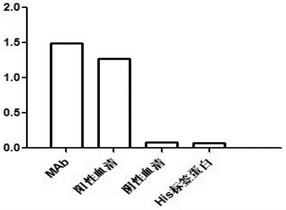
[0056] 1. Coating: The purified recombinant porcine epidemic diarrhea virus N protein and the pColdIDNA expression vector His-tagged protein were respectively coated on the ELISA microtiter plate with a coating volume of 200ng / 100μL per well, and coated overnight at 4°C.
[0057] 2. Washing: wash 3 times with PBST containing 5‰tween-20, 200 μL / well. 5 minutes each time.
[0058] 3. Blocking: Incubate with 5% skimmed milk at 37°C for 2 hours, 200 μL / well.
[0059] 4. Washing: wash 3 times with PBST containing 5‰ Tween-20, 200 μL / well. 5 minutes each time.
[0060] 5. Add primary antibody: Dilute the collected ascites with 5% skimmed milk at a ratio of 1:10000, add the enzyme label plate coated with ...
PUM
 Abstract
Description
Claims
Application Information
 - Generate Ideas
- Intellectual Property
- Life Sciences
- Materials
- Tech Scout
- Unparalleled Data Quality
- Higher Quality Content
- 60% Fewer Hallucinations
Browse by: Latest US Patents, China's latest patents, Technical Efficacy Thesaurus, Application Domain, Technology Topic, Popular Technical Reports.
© 2025 PatSnap. All rights reserved.Legal|Privacy policy|Modern Slavery Act Transparency Statement|Sitemap|About US| Contact US: help@patsnap.com