Three-dimensional nano-fiber tissue engineering scaffold and preparation method thereof

A fibrous tissue and three-dimensional nanotechnology, applied in the field of biomedicine, can solve the problems of low foreign gene capacity, short effective expression time, poor targeting specificity, etc., and achieve a good growth microenvironment, continuous stability, and high porosity Effect

Inactive Publication Date: 2016-04-20
ZHEJIANG MEDICAL COLLEGE
View PDF7 Cites 11 Cited by
  • Summary
  • Abstract
  • Description
  • Claims
  • Application Information

AI Technical Summary

Problems solved by technology

Viral vectors (such as various retroviruses, adenoviruses, poxviruses, herpesviruses, adeno-associated viruses, etc.) have high transfection efficiency in vivo, but there is a small capacity for exogenous genes (about 4.5-30kbp) , poor stability, poor targeting specificity, and potential toxicity

Method used


Image

  • Three-dimensional nano-fiber tissue engineering scaffold and preparation method thereof
  • Three-dimensional nano-fiber tissue engineering scaffold and preparation method thereof
  • Three-dimensional nano-fiber tissue engineering scaffold and preparation method thereof

Examples


preparation example Construction

[0027] The preparation method of the three-dimensional nanofibrous tissue engineering scaffold comprises:

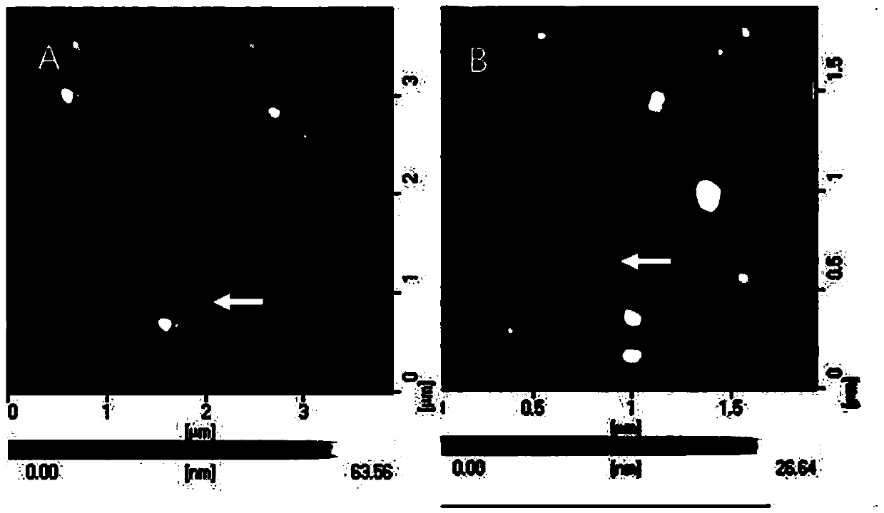
[0028] S1 Preparation of chitosan-nucleic acid nanocapsules

[0029] Specifically, firstly dissolve chitosan and nucleic acid in sodium acetate buffer solution respectively, the pH of sodium acetate solution is 5.4-5.6, and prepare 0.16-0.24 mg / mL chitosan solution and 0.08-0.12 mg / mL nucleic acid Solution, then heat the chitosan solution and the nucleic acid solution to 50-55°C respectively, then mix the chitosan solution and the nucleic acid solution, oscillate and mix the chitosan solution and the nucleic acid solution at a speed of 2400-2600rpm for 25-40s on a vortex oscillator, and then Stand still at room temperature for 30-60 minutes to obtain a chitosan-nucleic acid nano-microcapsule solution containing chitosan-nucleic acid nano-microcapsules. Preferably, the mass ratio of chitosan to nucleic acid in the obtained chitosan-nucleic acid nanocapsule solution is co...

Embodiment 1

[0051] First, prepare chitosan-nucleic acid nanocapsules

[0052] Chitosan and nucleic acid are dissolved in the 50mM sodium acetate buffer solution that pH is 5.5 respectively, in the present embodiment, used nucleic acid is plasmid pGPU6 / GFP / Neo (it can express green fluorescent protein in cell), certainly, can according to Select the appropriate nucleic acid for specific usage; prepare 0.2 mg / mL chitosan solution and 0.1 mg / mL pGPU6 / GFP / Neo solution; then heat the chitosan solution and pGPU6 / GFP / Neo solution to 50 ℃, then mix chitosan solution and pGPU6 solution according to the volume ratio of 1:1, mix well on the vortex shaker, 2500rpm, 30s; (pGPU6 / GFP / Neo) nanocapsule solution. Wherein, the mass ratio of chitosan to pGPU6 / GFP / Neo in the chitosan-nucleic acid (pGPU6 / GFP / Neo) nano-microcapsule solution is 2:1.

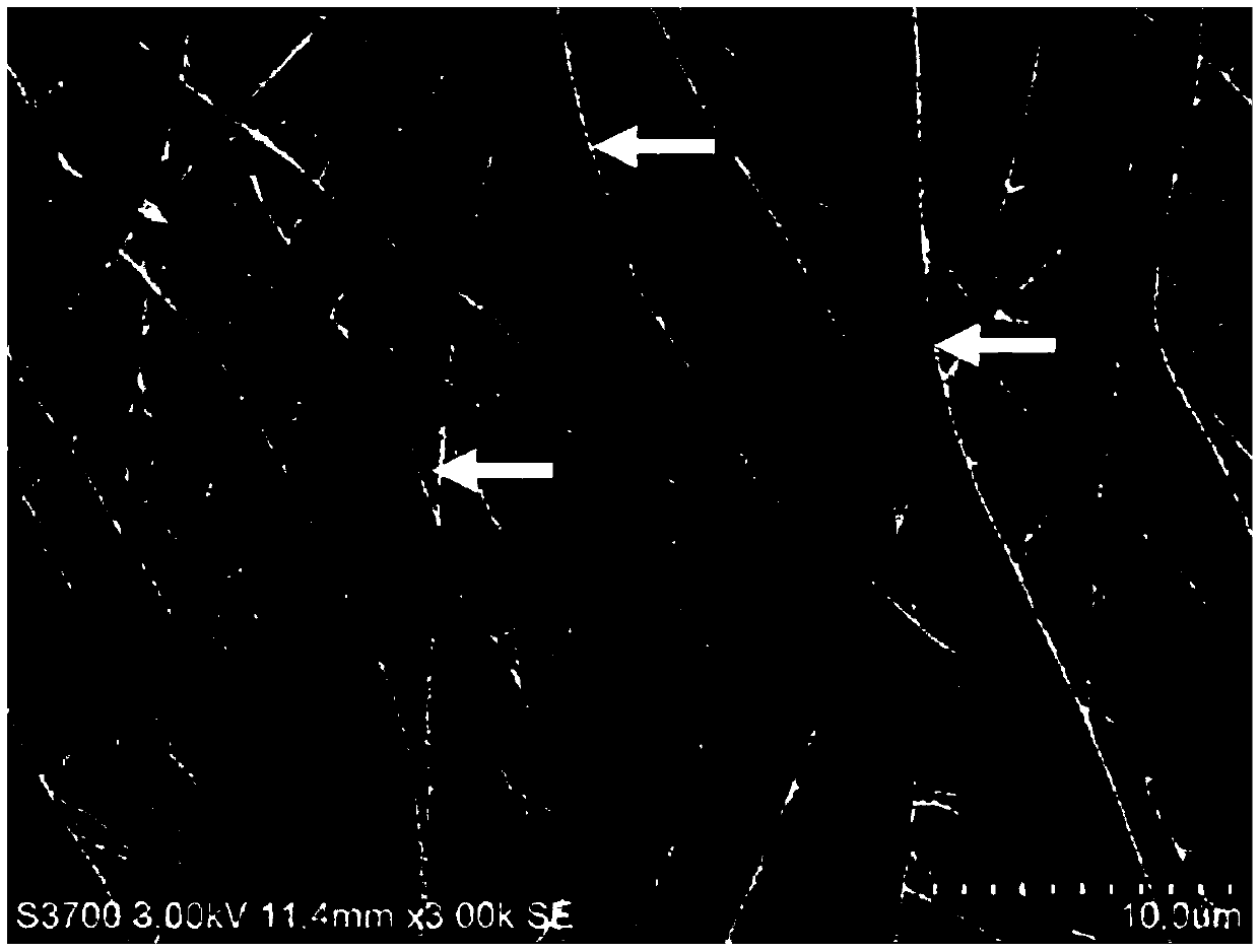
[0053] Secondly, configure the silk fibroin solution

[0054] The degummed silk fibroin is dissolved in the formic acid solution, stirred and mixed to completel...


PUM

PropertyMeasurementUnit
Particle sizeaaaaaaaaaa
Molecular weightaaaaaaaaaa

Abstract

The invention discloses a three-dimensional nano-fiber tissue engineering scaffold. The three-dimensional nano-fiber tissue engineering scaffold comprises a silk fibroin-chitosan-nucleic acid nano-fiber support which is formed by crosslinking silk fibroin-chitosan-nucleic acid nano-fiber filaments prepared from a spinning solution containing chitosan-nucleic acid nano micro-capsules and silk fibroin through a spinning process. The three-dimensional nano-fiber tissue engineering scaffold can provide a good growth micro-environment similar to an extracellular matrix and an enough mechanics support for cells and can be slowly and naturally degraded to release the chitosan-nucleic acid nano micro-capsules carrying nucleic acid, the chitosan-nucleic acid nano micro-capsules can guide carried nucleic acid into the cells, and the local targeting and time-controllable gene therapy effect is exerted. The invention further discloses a preparation method of the three-dimensional nano-fiber tissue engineering scaffold.

Description

technical field [0001] The invention relates to the field of biomedical technology, in particular to a three-dimensional nanofiber tissue engineering scaffold and a preparation method. Background technique [0002] Gene therapy is a biomedical treatment method that uses vectors to deliver target genes into target cells for moderate expression or interference for the purpose of treating diseases. Gene therapy consists of three basic elements: target gene, gene delivery carrier and target cells. Among them, how to obtain efficient, non-toxic, and biodegradable gene delivery vectors has become a technical bottleneck in the field of gene therapy. [0003] The vectors currently used in gene therapy can be divided into two categories: viral gene vectors and non-viral gene vectors. Viral vectors (such as various retroviruses, adenoviruses, poxviruses, herpesviruses, adeno-associated viruses, etc.) have high transfection efficiency in vivo, but there is a small capacity for exogen...

Claims


Application Information

Patent Timeline
IPC IPC(8): A61K47/48A61K48/00D04H1/728D01D5/00D01F4/00D01F1/10A61K47/61A61K47/62
CPCA61K48/00D01D5/0007D01F1/10D01F4/00D04H1/728
Inventor 银国利花扣珍傅晓斌蒋锦琴刘丹丹张琦
Owner ZHEJIANG MEDICAL COLLEGE
Why Eureka
Try Eureka