Fluorescence depletion method and microscopic imaging method and device

A fluorescence loss, microscopic imaging technology, applied in the field of optical microscopy, can solve the problems of super-resolution imaging loss wavelength scattering, and achieve the effect of reducing complexity, low price, and good optical properties

Active Publication Date: 2016-05-11
SOUTH CHINA NORMAL UNIVERSITY
View PDF5 Cites 6 Cited by
  • Summary
  • Abstract
  • Description
  • Claims
  • Application Information

AI Technical Summary

Problems solved by technology

[0005] Another object of the present invention is to provide a microscopic imaging method based on the above-mentioned fluorescence loss method, which combines the advantages of multiphoton super-resolution technology and up-conversion nanomaterials, and realizes that bot

Method used


Image

  • Fluorescence depletion method and microscopic imaging method and device
  • Fluorescence depletion method and microscopic imaging method and device
  • Fluorescence depletion method and microscopic imaging method and device

Examples


Embodiment 1

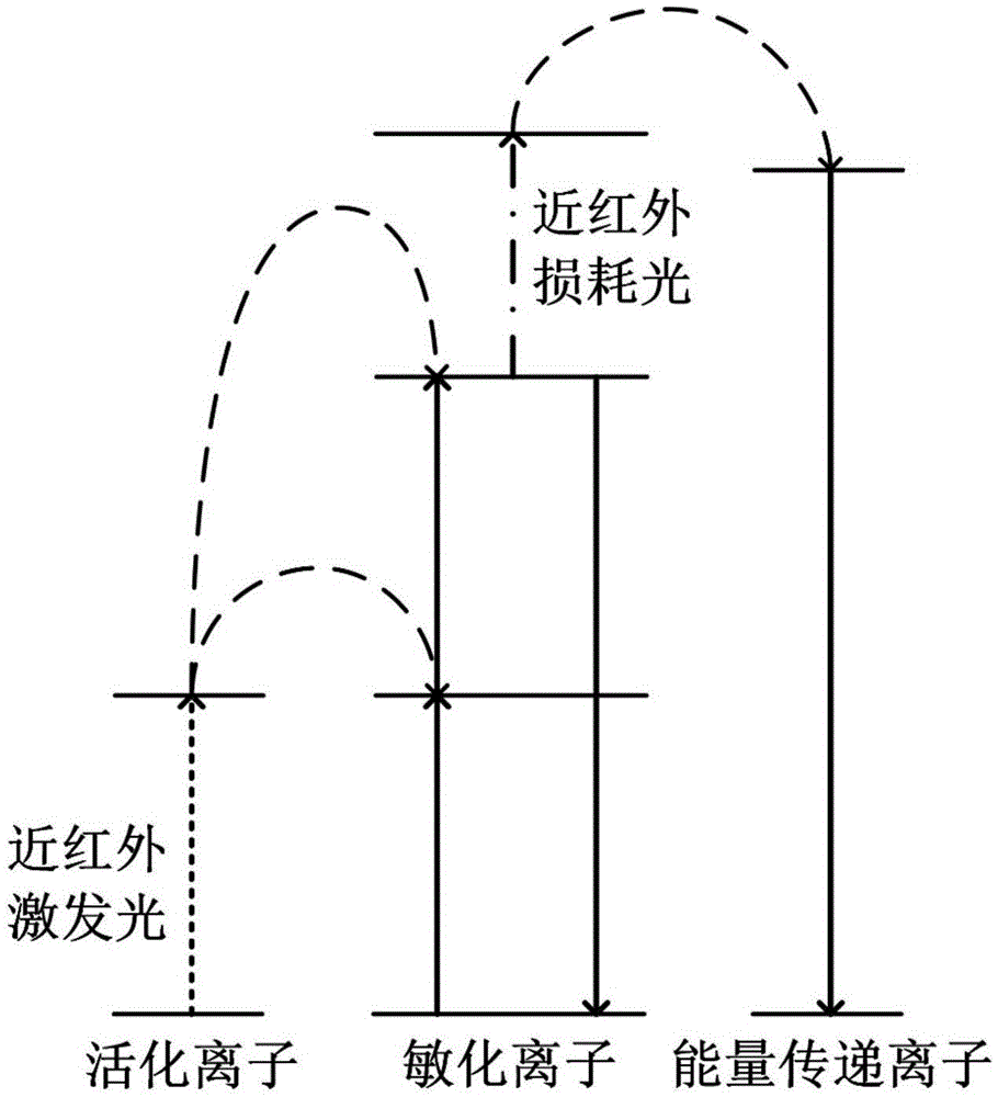
[0044] see figure 1 The wavelength bands of the near-infrared excitation light and the near-infrared depletion laser used in the fluorescence loss method in this embodiment are both between 760nm and 2000nm, and are used to excite rare earth-doped up-conversion nanomaterials. Rare earth ions in rare earth doped upconversion nanomaterials can be divided into three types according to their functions: activating ions, sensitizing ions and energy transfer ions. The steps are:

[0045] (1) Use near-infrared excitation light to excite rare earth-doped upconversion nanomaterials. After the activated ions absorb the near-infrared excitation light through ground state absorption, the energy is transferred to the sensitized ion through energy transfer upconversion and excited state absorption, and then The sensitized ions emit fluorescence in the ultraviolet, visible or near-infrared bands through the up-conversion process, that is, multi-photon fluorescence is excited.

[0046] (2) A...

Embodiment 2

[0052] Present embodiment except following feature other structures are with embodiment 1:

[0053] Such as Figure 5 shown, based on the upconversion NaYF 4 : Yb 3+ / Tb 3+ All two-photon fluorescence emitted under 980nm excitation (including 490nm, 546nm, 585nm, 620nm, etc.) can be consumed by another beam of near-infrared light (including 1108nm, 1264nm, 1374nm, 1541nm, etc.). In this example, Yb 3+ Acting as an activating ion, Tb 3+ As sensitizing ions and energy transfer ions.

[0054] The specific implementation process is as follows: under a certain power of 980nm laser excitation, two Yb 3+ After each ion absorbs a 980nm photon, a cooperative sensitization upconversion process (Cooperative Sensitization Upconversion, CSU) occurs, and it will be in the ground state 7 f 6 electrons excited to 5 D. 4 energy level. exist 5 D. 4 The electrons at the energy level will return to the corresponding low energy level by radiating fluorescence at 490nm, 546nm, 585nm, a...

Embodiment 3

[0056] Present embodiment except following feature other structures are with embodiment 1:

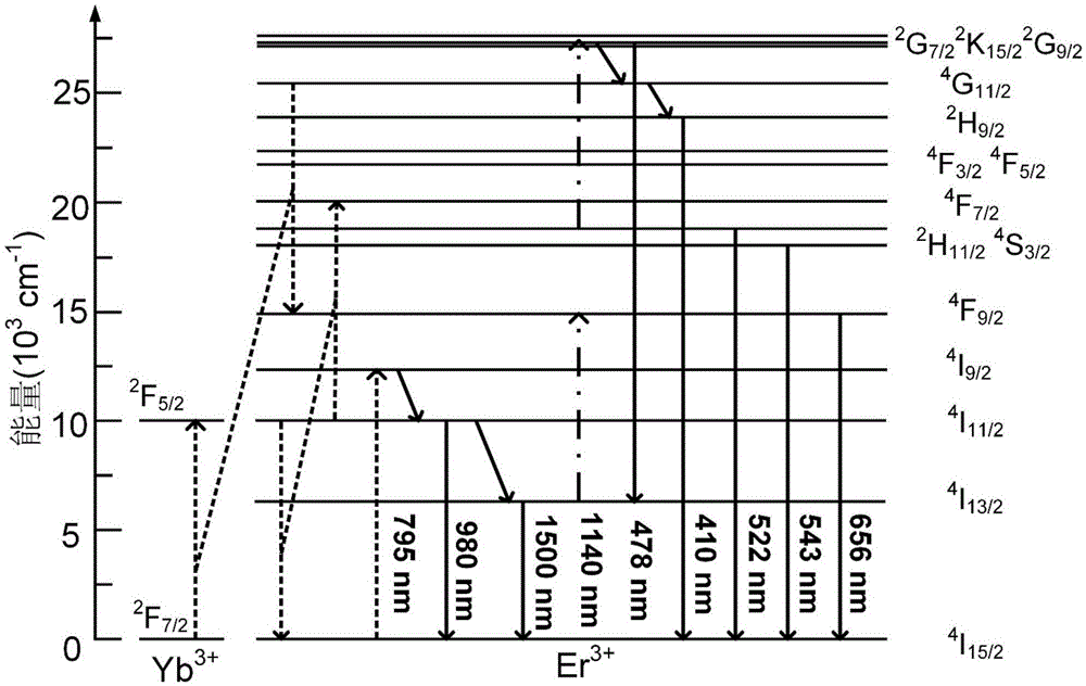
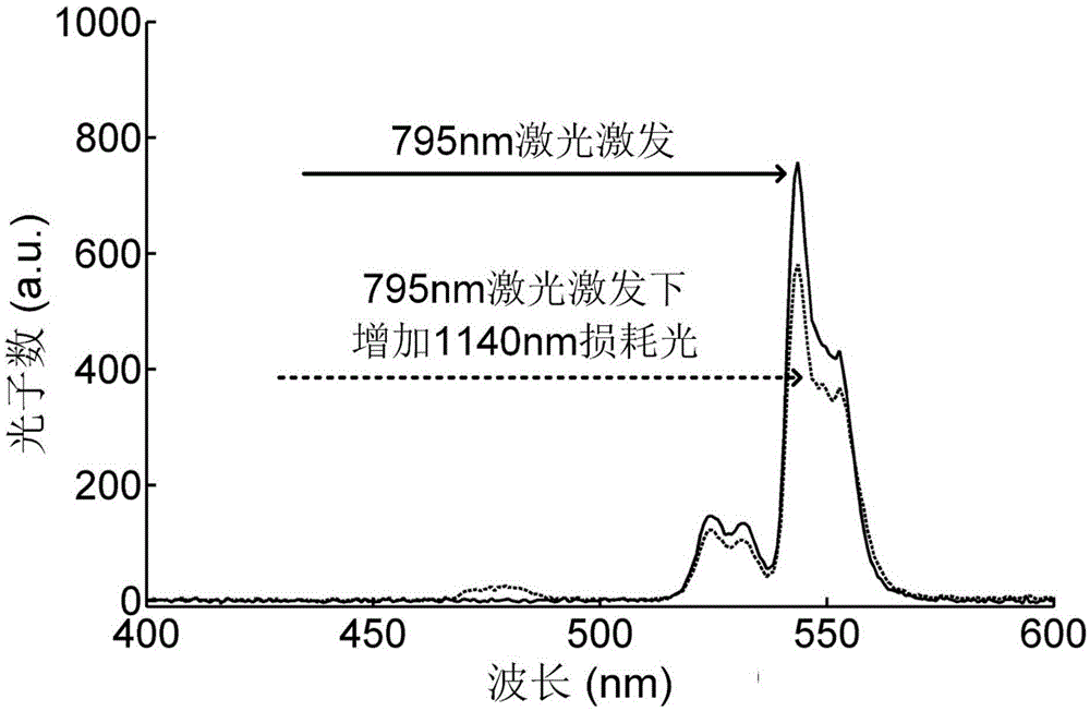
[0057] Based on the fluorescence loss method disclosed in Example 1, also based on up-conversion NaYF 4 : Yb 3+ / Er 3+ The green light emitted under 795nm excitation can be lost by 1140nm light. This embodiment provides a microscopic imaging method, the method comprising:

[0058] In the same way, a continuous laser with a wavelength of 795nm is used to emit a stable near-infrared wavelength laser as the excitation light. After the laser is filtered by a collimating beam expander and a small aperture diaphragm, a focused Gaussian solid spot is obtained;

[0059] At the same time, in the other path, a continuous laser with a wavelength of 1140nm is used to generate a stable near-infrared wavelength laser as a near-infrared loss laser. Modulate and form a hollow beam to obtain a stimulated emission depletion spot; the wavelength of the near-infrared depletion laser conforms to the 2...


PUM

PropertyMeasurementUnit
The average diameteraaaaaaaaaa

Abstract

The invention discloses a fluorescence depletion method and a microscopic imaging method and device. The fluorescence depletion method is characterized in that near-infrared depletion light is used to cause stimulated absorption in the sensitizing ions of rare earth doped upconversion nano materials, the energy of near-infrared excitation light is transferred to energy transfer ions, and depletion of the multiphoton fluorescence of the upconversion nano materials is achieved. The microscopic imaging method is provided on the basis of the fluorescence depletion method, and the microscopic imaging method is characterized in that the near-infrared depletion light is modulated into a hollow light beam by using a space phase modulation plate, and the hollow light beam and an excitation light beam are subjected to collimation conjugation focusing to achieve microscopic imaging of the rare earth doped upconversion nano materials and the marked samples of the rare earth doped upconversion nano materials. A super-resolution optical microscopic imaging system formed by an excitation light generating module, a depletion light generating module, a dichroscope, a multiphoton microscopic scanning module and a photoelectric detection module is built on the basis of the microscopic imaging method. By the fluorescence depletion method and the microscopic imaging method and system, low-cost, low-complexity, high-resolution, simple and effective real-time dynamic three-dimensional images can be obtained.

Description

technical field [0001] The invention belongs to the field of optical microscopy technology, and in particular relates to a fluorescence loss method for realizing double-near-infrared wavelength excitation, and a microscopic imaging method and a microscopic imaging device using the fluorescence loss method. Background technique [0002] In the conventional optical imaging process, according to Abbe's principle, the limit resolution that the optical system can achieve is about half of the wavelength of the incident light. In order to improve the resolution, scientists have proposed many methods to break the diffraction limit, collectively referred to as super-resolution imaging methods. One of the important methods is stimulated radiation depletion (STimulatedEmissionDepletion, STED). STED microscopy consists of an excitation beam and a phase-modulated hollow beam. In STED, the method of stimulated radiation is used to forcibly quench the fluorescence at the periphery of the...

Claims


Application Information

Patent Timeline
IPC IPC(8): G01N21/64
CPCG01N21/6458
Inventor 詹求强吴锐涛王保举
Owner SOUTH CHINA NORMAL UNIVERSITY
Why Eureka
Try Eureka