Preparation method and application of angiogenesis model

An angiogenesis and model technology, applied in the field of biomedicine, to achieve the effect of good development and application prospects

Inactive Publication Date: 2016-07-27
WUHAN UNIV
View PDF1 Cites 3 Cited by
  • Summary
  • Abstract
  • Description
  • Claims
  • Application Information

AI Technical Summary

Problems solved by technology

However, the traditional model of angiogenesis lacks the participation of type Ⅰ collagen, so the traditional model of matrigel angiogenesis cannot accurately reflect the abnormal growth of blood vessels in the synovium during TMJOA.
In addition, in numerous studies, ECs were not observed for more than 24 hours after inoculation

Method used


Image

  • Preparation method and application of angiogenesis model
  • Preparation method and application of angiogenesis model
  • Preparation method and application of angiogenesis model

Examples


Embodiment 1

[0027] [Example 1] Culture of umbilical vein endothelial cells

[0028] Umbilical vein vascular endothelial cells (HUVECs) were purchased from Nanjing Kaiji Biotechnology Co., Ltd., the article number is KG-110, and the umbilical vein vascular endothelial cells (HUVECs) containing 10% fetal bovine serum (fetalbovineserum, FBS, SV30087, ) in RPMI-1640 medium. Cells were digested using 0.25% trypsin (EDTA free).

Embodiment 2

[0029] [Example 2] Construction of traditional matrigel angiogenesis model

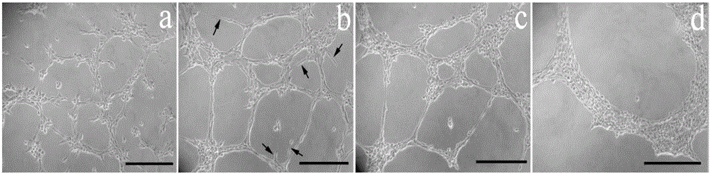
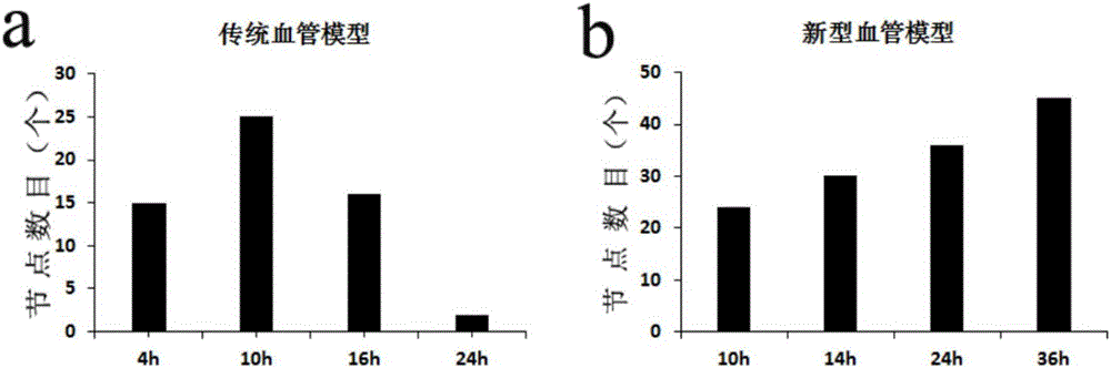
[0030] in 24-well plate Add 150 μl of liquid Matrigel (356230, BD Biosciences, Bedford, MA, USA) at 4°C to the cell culture medium, and put it in a constant temperature cell incubator at 37°C for 20 minutes to make it change from a liquid state to a gel state. Digest HUVECs and replace with vascular endothelial cell-specific medium containing 10% FBS (CC-3156, Lonza, Walkersville, MD, USA) neutralize the digestion reaction, count, add appropriate medium to make 0.5ml Contains 5x10 4 HUVECs were seeded on the surface of Matrigel, and the 24-well plate was placed in an environment containing 95% air and 5% CO 2in a constant temperature cell culture incubator at 37°C. Use a phase contrast microscope to observe and take pictures at different time points, and calculate the number of nodes in the capillary network structure at each time point. The method is: each 24-well plate is observed with a 40...

Embodiment 3

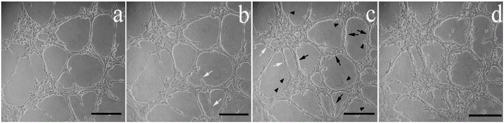
[0032] [Example 3] Construction of a new angiogenesis model

[0033] The preparation process of type Ⅰ rat tail collagen is as follows: Add 200 μl type Ⅰ rat tail collagen (Shengyou Biotechnology Co., Ltd.) into a 50ml centrifuge tube placed in ice, add 690 μl double distilled water, mix well, add to another In a 50ml centrifuge tube, 12μl of 0.1mol / l NaOH has been added to it, and immediately mixed quickly. Add 100 μl 10x phosphate buffered saline (PBS) and mix well. The whole process was placed on ice, and the preparation of neutralized 1 mg / ml type I rat tail collagen was completed at this time.

[0034] This model is based on the traditional matrigel angiogenesis model mentioned above, that is, when HUVECs were inoculated on matrigel for 12 hours, all the culture medium on the surface of matrigel was discarded, and 400 μl of liquid neutralized type I rat tail collagen was added. Place the 24-well plate in a constant temperature cell culture incubator at 37°C for 20 minut...


PUM


Abstract

The invention provides a preparation method and application of an angiogenesis model. The method includes the following steps that firstly, vascular endothelial cells are inoculated to matrigel to primarily form vasoganglion; secondly, type I collagen is added to stimulate secondary angiogenesis. The preparation method is suitable for simulating the pathogenesis process of angiogenesis in joint synovial tissue and particularly suitable for simulating and building angiogenesis conditions in temporo-mandibular joint synovial tissue of a human body, and provides a new platform for research of action modes of new cytokines in the aspect of angiogenesis promotion or inhibition. The model is simple in manufacturing material and short in consumed time and has good development, expansion and tissue engineering application prospects.

Description

technical field [0001] The invention belongs to the field of biomedicine, and in particular relates to a preparation method of a novel angiogenesis model. Background technique [0002] Temporomandibular joint (TMJ) osteoarthritis (OA) is a degenerative disease characterized by the destruction and wear of articular cartilage, accompanied by changes in subchondral bone tissue and corresponding changes in the synovial membrane. The main clinical manifestations are joint area pain, murmur and abnormal jaw movement. During the pathogenesis of TMJOA, chronic inflammation in the synovial tissue often recurs, and hyperemia of the synovial tissue can be seen under arthroscopy. Inflammation and angiogenesis complement each other: in the inflammatory phase, synoviocytes lose the components that protect and nourish cartilage, such as hyaluronic acid and lubricant, and secrete various inflammatory factors, such as IL-1β, IL-6, TNF-α, VEGF, HMGB1, MMPs, etc., promote angiogenesis, degra...

Claims


Application Information

Patent Timeline
IPC IPC(8): C12N5/071C12Q1/02
CPCC12N5/0691C12N2503/02C12N2533/54G01N33/5088G01N2800/7014
Inventor 李颖杰龙星
Owner WUHAN UNIV
Why Eureka
Try Eureka