Spontaneously immortalized piglet oral mucosa epithelial cell line and construction method thereof

A technology of oral mucosal epithelium and establishment method, applied in the field of spontaneously immortalized piglet oral mucosal epithelial cell line and its establishment, can solve the problems of limited number of passages, limited application, poor cell stability and uniformity, etc., and achieve rapid and stable growth Good performance and stable performance

Inactive Publication Date: 2016-08-10
张彦明
View PDF2 Cites 2 Cited by
  • Summary
  • Abstract
  • Description
  • Claims
  • Application Information

AI Technical Summary

Problems solved by technology

[0004] The object of the present invention is to provide a kind of spontaneously immortalized piglet oral mucosal epithelial cell line and its establishment method, aiming at solving the problem that the number of passages of oral mucosal epithelial cells in in vitro culture is limited, and the cell stability and uniformity are very poor, which greatly limits the problems with its application

Method used


Image

  • Spontaneously immortalized piglet oral mucosa epithelial cell line and construction method thereof
  • Spontaneously immortalized piglet oral mucosa epithelial cell line and construction method thereof
  • Spontaneously immortalized piglet oral mucosa epithelial cell line and construction method thereof

Examples


Embodiment 1

[0035] Example 1 Primary culture of piglet oral mucosal epithelial cells

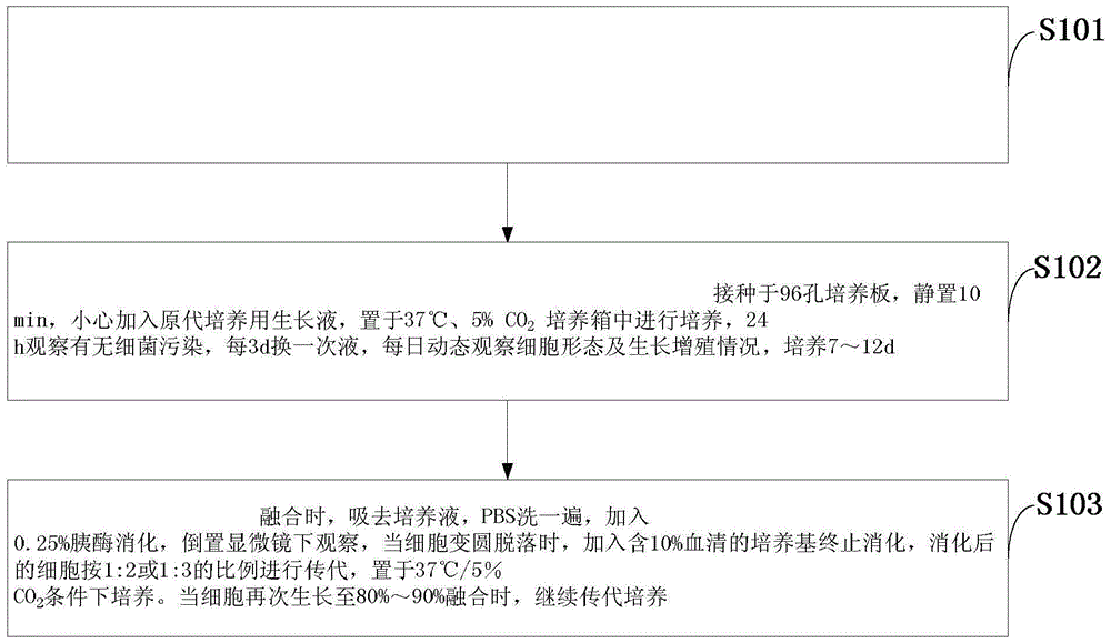
[0036] After the non-lactating piglets were anesthetized, blood was collected from the heart to kill them, disinfected with alcohol, and the oral epithelial tissues corresponding to the cheeks were cut, rinsed several times with PBS (containing 600IU / ml double antibody, 5μg / mL amphotericin B) under sterile conditions ,, remove the non-mucosal epithelial tissue visible to the naked eye, cut the oral mucosal epithelial tissue into 0.2cm×1cm tissue pieces, transfer them into centrifuge tubes, add 2.5mg / mL DispaseII solution, digest at 4°C for 18-20h, and then cut the tissue Into 3 Small pieces of tissue, rinsed 3 times with PBS, centrifuged at 1000r / min for 5min, inoculated in 96-well culture plate, let stand for 10min, carefully added growth medium for primary culture, and placed at 37°C, 5% CO 2 Cultivate in an incubator, observe for 24 hours whether there is bacterial contamination, change the liquid ev...

Embodiment 2

[0037] Example 2. Subculture of piglet oral mucosal epithelial cells

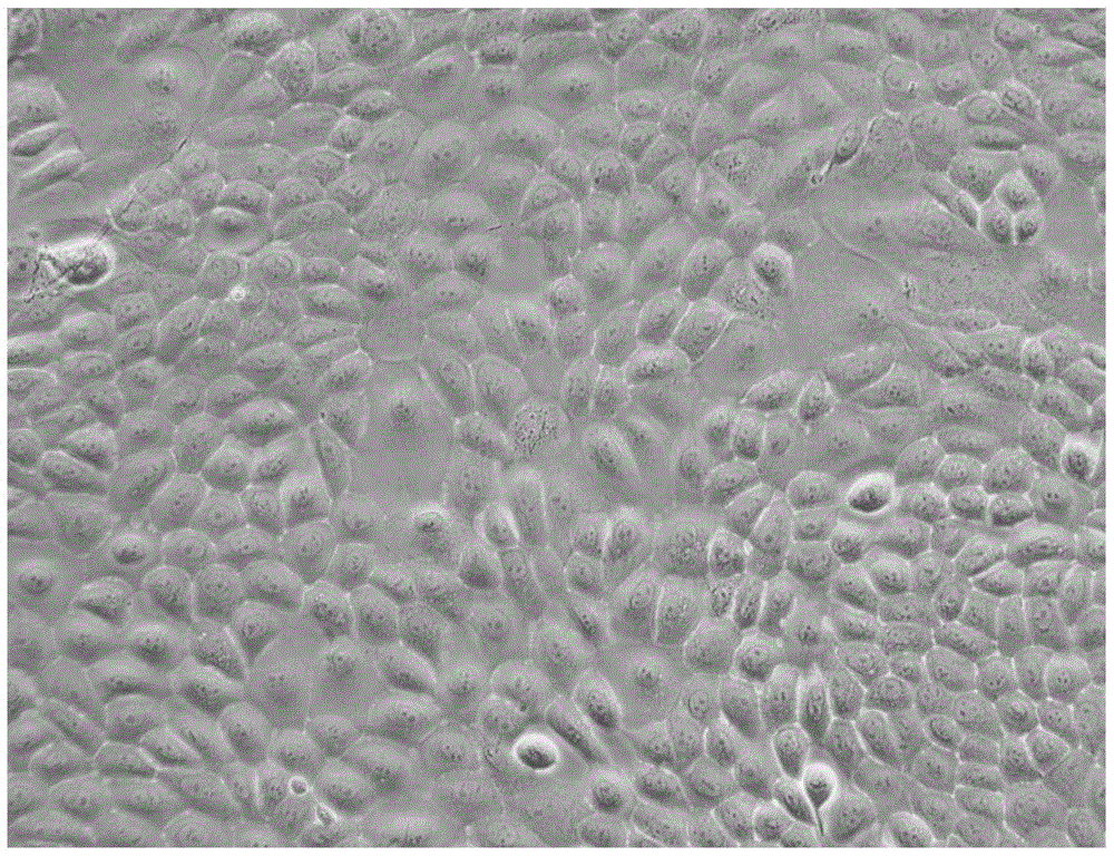
[0038] When the primary cells grow to 80% to 90% confluence, remove the culture medium, wash with PBS, add 0.25% trypsin to digest, observe under an inverted microscope, when the cells become round and fall off, add the medium containing 10% serum to terminate Digestion, the digested cells are passaged at a ratio of 1:2 or 1:3, and placed at 37°C / 5%CO 2 cultivated under conditions. When the cells grow to 80%-90% confluence again, continue to subculture in this way. The cells have been passaged for more than 100 passages ( image 3 ), the cells still maintain the characteristics of normal epithelial cells.

Embodiment 3

[0039] Example 3. Identification of Cell Types by Indirect Immunofluorescence

[0040]1. Plating: Place a sterile coverslip of 0.5 cm × 0.6 cm in the well of a 24-well cell culture plate, digest piglet oral mucosa epithelial cells into single cells with trypsin, and adjust the cell concentration to 2 × 10 after counting. 5 cells / mL, inoculate 0.5 mL of cell suspension in each well.

[0041] 2. Fixing: When the cells grow to a near-confluent state (low-density monolayer), wash with PBS (0.01mol / L, pH 7.4) 3 times, add 0.4g / L paraformaldehyde phosphate buffer (pH 7.4), Cells face up, fixed at room temperature for 10 min, washed 3 times with PBS, 3 min each time.

[0042] 3. Breakthrough: add 1% Triton breakthrough solution (prepared in PBS), act at 37°C for 15 minutes, wash with PBS 3 times, 3 minutes each time.

[0043] 4. Blocking: 5% skimmed milk powder in PBS solution, 37° C., blocking for 1 hour.

[0044] 5. Incubate the primary antibody: Shake off the blocking solution,...


PUM


Abstract

The invention discloses a spontaneously immortalized piglet oral mucosa epithelial cell line and a construction method thereof. Oral mucosa epithelial tissue of a newly-born unfed piglet is subjected to primary culture and continuous passage culture so that a high-purity and good-stability spontaneously immortalized piglet oral mucosa epithelial cell line is obtained. The spontaneously immortalized piglet oral mucosa epithelial cell line can fast grow and has stable performances. 110th generation of cells still keep form characteristics of primary cells. A karyotype analysis result shows that the cells do not produce mutation. The spontaneously immortalized piglet oral mucosa epithelial cell line is self-formed after serial passage, does not produce exogenous gene insertion and gene mutation, can be used as a basic cell in vaccine research and development and does not produce a biological safety risk. The cell line provides a good cell model for molecular biology and virology research and can be used for porcine virus vaccine research and development.

Description

technical field [0001] The invention belongs to the technical field of cell biology, and in particular relates to a spontaneously immortalized piglet oral mucosa epithelial cell line and a method for establishing the same. Background technique [0002] Under natural conditions, pathogens such as viruses and bacteria invade hosts mostly through the oral and nasal route. Oral mucosal epithelial cells, as the first line of defense of the animal immune system, are the earliest cells encountered by viruses and bacteria infected hosts. Oral mucosal epithelial cells express a variety of pattern recognition receptors, which can recognize pathogen-associated molecular patterns expressed by most invading pathogens. Oral mucosal immunity is an important part of the body's innate immunity. [0003] There are great difficulties in the in vitro culture of oral mucosal epithelial cells, the number of passages is limited, and the stability and uniformity of the cells are poor, which greatl...

Claims


Application Information

Patent Timeline
Patent Type & Authority Applications(China)
IPC IPC(8): C12N5/071C12R1/91
CPCC12N5/0632C12N2509/00C12N2509/10
Inventor 张彦明崔红杰郭抗抗
Owner 张彦明
Why Eureka
Try Eureka