Biofilm capable of rapidly recovering skin wound surface, and preparation method thereof
A skin wound, rapid recovery technology, applied in biochemical equipment and methods, microorganisms, skin diseases, etc., can solve the problems of high cost, unfavorable promotion and application, slow effect, etc., to achieve low cost, reduce deterioration, and protect wounds.
- Summary
- Abstract
- Description
- Claims
- Application Information
AI Technical Summary
Problems solved by technology
Method used
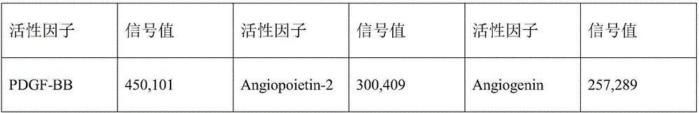
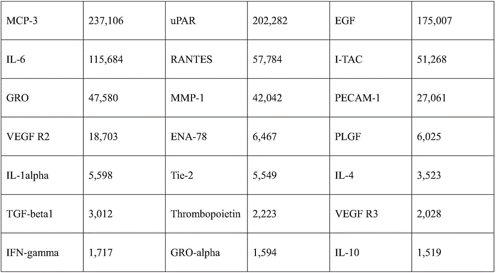
Image

Examples
Embodiment 1
[0021] Example 1 EPC cell culture fluid calcium alginate composite biofilm (Ⅰ)
[0022] 1) Extraction of human umbilical cord blood endothelial progenitor cells: extract 60ml of umbilical cord blood from healthy newborns with a syringe, anticoagulate with sodium heparin, dilute with PBS at a volume ratio of 1:1, and slowly add Ficoll (1.077) lymphocyte separation solution containing a volume ratio of 2:1 In the centrifuge tube, use the density gradient centrifugation method, centrifuge at 2000rpm in a horizontal centrifuge for 25 minutes, and carefully absorb the mononuclear cell layer with a pipette. Then wash the cells with an equal volume of PBS, centrifuge at 2000rpm for 8 minutes, and inoculate the cells on a 25cm cell coated with human fibronectin. 2 Add an appropriate amount of endothelial cell culture medium (containing 10% fetal bovine serum) to the culture flask for culture, and culture at a saturated humidity, 5% carbon dioxide concentration, and a constant temperat...
Embodiment 2
[0027] Example 2 EPC cell culture fluid calcium alginate composite biofilm (Ⅱ)
[0028] The endothelial progenitor cells were subcultured according to the steps of 1) and 2) in Example 1. After culturing the cells to the third generation, when the cells grow to 70-80% confluence, place the cells in 0.5% oxygen concentration, saturated humidity, and 5% carbon dioxide concentration conditions to induce hypoxia for 36 hours, after that, replace the cell culture medium with no The serum basal culture medium was restored to 21% oxygen concentration, and cultured for 72 hours under saturated humidity and 5% carbon dioxide concentration. After centrifugal filtration, the culture medium was collected, and the calcium alginate composite biofilm was prepared by freeze-drying method.
Embodiment 3
[0029] Example 3 EPC cell culture fluid calcium alginate composite biofilm (Ⅲ)
[0030] The endothelial progenitor cells were subcultured according to the steps of 1) and 2) in Example 1. After culturing the cells to the 5th generation, when the cells grow to 70-80% confluence, place the cells in 1.5% oxygen concentration, saturated humidity, and 5% carbon dioxide concentration conditions to induce hypoxia for 24 hours, after that, replace the cell culture medium with no The serum basal culture solution was restored to 21% oxygen concentration, and cultured for 48 hours under saturated humidity and 5% carbon dioxide concentration. After centrifugal filtration, the culture medium was collected, and the calcium alginate composite biofilm was prepared by freeze-drying method.
PUM
 Abstract
Description
Claims
Application Information
 - R&D
- Intellectual Property
- Life Sciences
- Materials
- Tech Scout
- Unparalleled Data Quality
- Higher Quality Content
- 60% Fewer Hallucinations
Browse by: Latest US Patents, China's latest patents, Technical Efficacy Thesaurus, Application Domain, Technology Topic, Popular Technical Reports.
© 2025 PatSnap. All rights reserved.Legal|Privacy policy|Modern Slavery Act Transparency Statement|Sitemap|About US| Contact US: help@patsnap.com