
Method for extracting bacterial outer membrane vesicles

A technology for outer membrane vesicles and membrane vesicles, which is applied in the field of preparing Escherichia coli and Klebsiella pneumoniae efflux membrane vesicles, can solve problems such as troublesome handling and complicated operation, and achieve the effect of convenient operation

Inactive Publication Date: 2017-06-09
FUDAN UNIV
View PDF4 Cites 3 Cited by
  • Summary
  • Abstract
  • Description
  • Claims
  • Application Information

AI Technical Summary

Problems solved by technology

[0005] The current extraction methods for OMVs mainly include sucrose density gradient centrifugation and Optiprep density gradient centrifugation. However, practice shows that these two methods are cumbersome to operate and the post-processing is troublesome.

Method used


Image

  • Method for extracting bacterial outer membrane vesicles
  • Method for extracting bacterial outer membrane vesicles
  • Method for extracting bacterial outer membrane vesicles

Examples


Embodiment 1

[0026] Embodiment 1: Selection and cultivation of bacterial strains

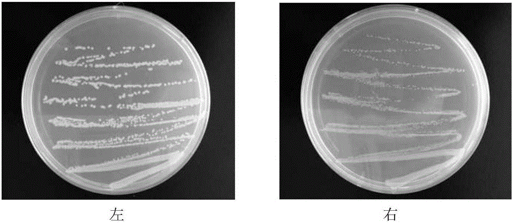
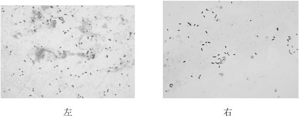
[0027] Klebsiella pneumoniae was purchased from the China Agricultural Microorganism Culture Collection, and Escherichia coli DH5α was purchased from the American Culture Collection. Klebsiella pneumoniae was cultured with NB medium, and Escherichia coli was cultured with LB medium; the freeze-dried powder of the strains was dissolved in sterile saline, and an appropriate amount was placed on a solid medium, and cultured overnight at 37°C until a single colony (such as figure 2 shown); single colonies were cultured in test tubes containing 3ml of liquid medium, and cultured overnight at 37°C on a shaker at 180r / min. The next day, transfer 3ml of the bacterial solution to a shake flask containing 40ml of medium for cultivation until the OD value of the bacteria reaches 1.0. image 3 The cell morphology after crystal violet staining is shown.

Embodiment 2

[0028] Embodiment 2: Extraction of OMVs

[0029] Centrifuge the bacterial solution cultivated to an OD value of 1.0 in a 50ml sterile centrifuge tube at 6000r / min for 20min, remove most of the bacterial cells, filter the supernatant with a 0.22μM sterile filter head to completely sterilize, and then transfer the filtrate Centrifuge at 3000r / min for 10min at 4°C for 10min in 50ml of ultrafiltration tube with a molecular weight cut-off of 100KD, and repeat several times until the solution is overall and concentrated to about 1 / 8 of its original volume;

[0030] Add 5ml of normal saline to the concentrated solution, use the ultrafiltration concentration method to wash away impurities such as excess protein and flagella pili, dispense it into EP tubes, and store at -20°C.

Embodiment 3

[0031] Example 3: Identification of OMVs

[0032] A, BCA method to the determination of OMVs protein concentration;

[0033] OMVs protein concentration was measured by BCA method;

[0034] Take 5 mg / ml standard protein and establish protein-absorbance standard curve in 96-well plate (such as Figure 4 As shown), take 2 μl OMVs samples in the other 5 auxiliary wells, add 200 μl AB solution (A solution: B solution = 50:1) in each well, incubate in a 37°C incubator for 30min, measure the absorbance value of each well at a wavelength of 570nm ; To establish a standard curve for protein content, R 2 =0.9989>0.99, showing a good linear relationship;

[0035] The measured protein concentration of Klebsiella pneumoniae OMVs is about 1.5 mg / ml, and the concentration of Escherichia coli OMVs is about 2.1 mg / ml;

[0036] Table 1 shows the protein concentration of the extracted Klebsiella pneumoniae and Escherichia coli OMVs.

[0037] Table 1

[0038]

[0039] B. Negative stain ele...


PUM

PropertyMeasurementUnit
Molecular weightaaaaaaaaaa

Abstract

The invention discloses a method for obtaining outer membrane vesicles (OMVs) of escherichia coli and klebsiella pneumoniae by utilizing an ultrafiltration and concentration method. The method comprises the following steps of making the escherichia coli and the klebsiella pneumoniae into bacterial suspension, taking and inoculating a proper amount of the bacterial suspension into a test tube containing a culture medium for culturing and staying overnight, then taking and inoculating a proper amount of a bacterial solution into a shake flask containing a culture medium l for culturing until an OD (Optical Density) value approaches 1.0, collecting the bacterial solution, centrifuging the bacterial solution to remove a thallus, filtering the bacterial solution by a sterile filter head to remove a residual thallus, collecting filtrate, centrifugally concentrating the filtrate until 1 / 8 of an original volume by an ultrafiltration tube with a molecular weight cutoff of 100KDa, adding normal saline into the filtrate for washing, repeating the step for 2 to 3 times, collecting the bacterial outer membrane vesicles located on an upper layer of the ultrafiltration tube, and putting the bacterial outer membrane vesicles in an environment at subzero 20 DEG C for subpackaging and cryopreserving, wherein the protein concentration of the extracted outer membrane vesicle of the klebsiella pneumoniae is 1.5mg / ml; the protein concentration of the extracted outer membrane vesicle of the escherichia coli is 2.1mg / ml; the particle sizes of the OMVs are approximately 20nm to 100nm. The method is convenient, simple, easy and feasible to operate; a novel method is provided for the extraction and the research of the OMVs.

Description

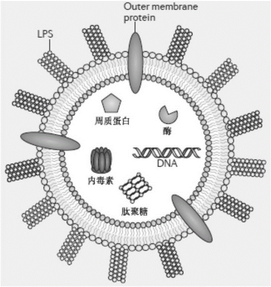
technical field [0001] The invention relates to a method for preparing escherichia coli (DH5α) and Klebsiella pneumoniae (klebsiella pneumoniae, KP) efflux membrane vesicles (bacterial outer membrane vesicles, OMVs) by using an ultrafiltration concentration method. Background technique [0002] Bacterial outer membrane vesicles are reported to be bilayered spherical proteoliposomes that are continuously released in the form of budding from the surface of Gram-negative bacteria, generally 20–250 nm in diameter, and usually carry bacterial outer membrane and periplasmic components, including Some DNA, RNA, endotoxin (LPS), protein, enzyme and peptidoglycan etc., its structure is as figure 1 shown; clinical studies have shown that OMVs can cause inflammation and pathological responses in the body, and OMVs are associated with many infectious diseases, such as periodontitis, gastritis, Crohn's disease, salpingitis, meningitis, sepsis, cardiovascular disease As well as lung dise...

Claims


Application Information

Patent Timeline
IPC IPC(8): C12N1/20A61K47/46A61K39/02A61P31/04C12R1/19C12R1/22
CPCA61K39/0258A61K39/0266A61K47/46C12N1/20
Inventor 叶丽姜珊珊王乾冯美卿
Owner FUDAN UNIV
Learn More