Fluorescent nanoprobe, preparation method thereof and application thereof in biosensor

A fluorescent nanoprobe and nanomaterial technology, applied in biosensing applications, fluorescent nanoprobes and their preparation fields, can solve problems such as cell or tissue damage, toxicity, and difficulty in metabolism, and achieve simple preparation methods, cellular Low toxicity, accurate qualitative and quantitative effects

Active Publication Date: 2017-06-30
NANJING UNIV
View PDF5 Cites 6 Cited by
  • Summary
  • Abstract
  • Description
  • Claims
  • Application Information

AI Technical Summary

Problems solved by technology

However, when the above-mentioned two-dimensional nanomaterials are applied to intracellular or in vivo analysis, they will encounter the problem of difficulty in metabolism, and then produce certain toxicity, causing damage to cells or tissues.

Method used


Image

  • Fluorescent nanoprobe, preparation method thereof and application thereof in biosensor
  • Fluorescent nanoprobe, preparation method thereof and application thereof in biosensor
  • Fluorescent nanoprobe, preparation method thereof and application thereof in biosensor

Examples


Embodiment 1

[0042] 1: Preparation of MOF-La crystal

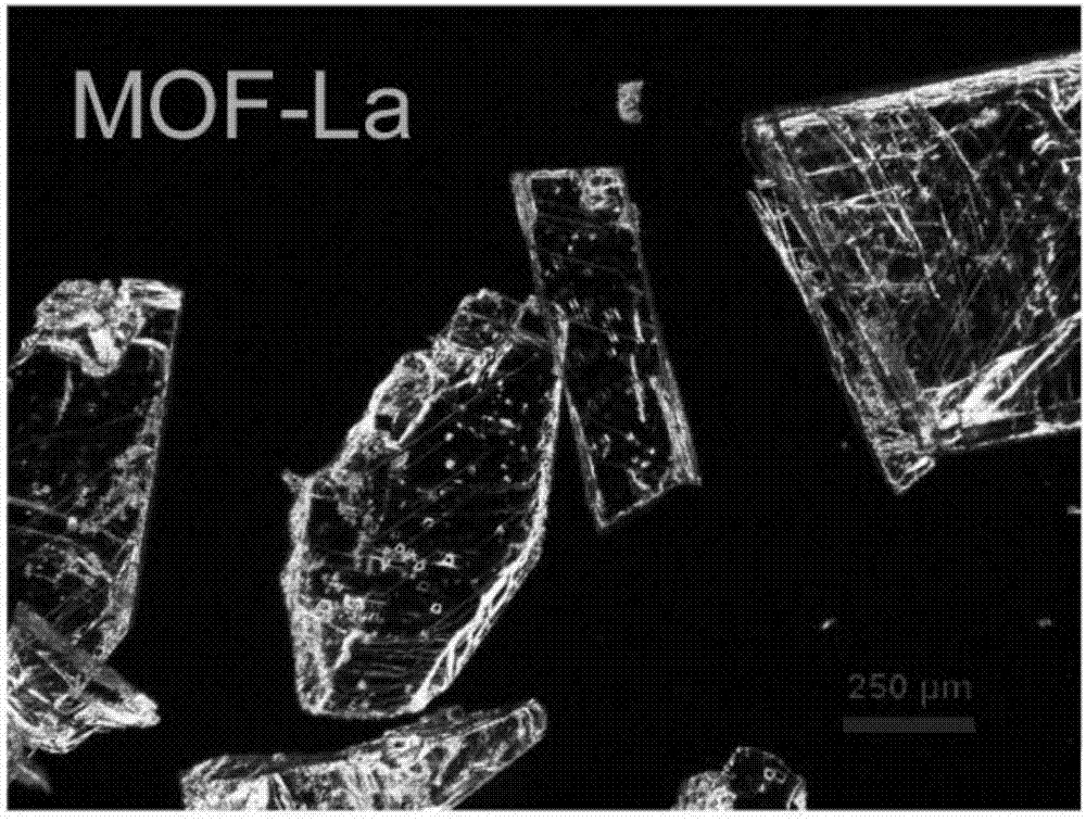
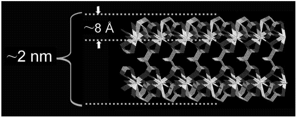
[0043] (1) In 15 mL of NaOH solution (100 mM), add 120 mg (0.8 mmol) of 2,2′-thiodiacetic acid and dissolve by ultrasonication. Then add 173 mg (0.4 mmol) La(NO 3 ) 3 ·6H 2 O, shake to dissolve. The obtained solution was transferred to the polytetrafluoroethylene liner of the reaction kettle, the reaction kettle was sealed, put into an oven, heated at 150°C, and reacted for 15 h. After the reaction, the reactor was cooled down to room temperature to obtain MOF-La crystals, whose morphology was as follows: figure 1 shown. structured as figure 2 shown.

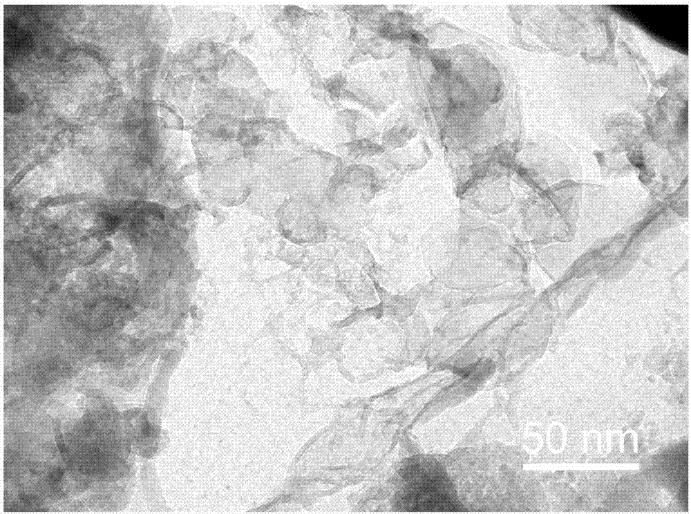
[0044](2) Add the MOF-La (5 mg) obtained from the above reaction into 2 mL of ethanol, sonicate for 30 min with a cell disruptor, and centrifuge at 8000 rpm to obtain the initially stripped MOF-La. Then the MOF-La was dispersed in 10 mL of n-butyllithium hexane solution (0.16 M), under nitrogen protection, and stirred for 20 h at 25 °C to obtain the exfoliated two-dimensional M...

Embodiment 2

[0053] (1) In 15 mL KOH solution (70 mM), add 77 mg (0.45 mmol) 2,2′-sodium thiodiacetate, dissolve and add 130 mg (0.225 mmol) La 2 (SO 4 ) 3 dissolve. Then heated at 120°C and reacted for 10 h. After the reaction, the temperature was lowered to room temperature to obtain MOF-La crystals.

[0054] (2) Add MOF-La (5 mg) obtained from the above reaction into 2 mL of n-hexane, sonicate for 35 min with a cell disruptor, and then centrifuge to obtain MOF-La that has been preliminarily stripped.

[0055] (3) Disperse the preliminarily stripped MOF-La in 10 mL of n-butyllithium cyclohexane solution (0.16 M), protect it with helium, and stir the reaction for 25 h at 30°C to obtain the stripped Two-dimensional MOF-La.

[0056] (4) Disperse 0.2 mg of two-dimensional MOF-La in 1 mL of PBS (0.01 M, pH 8.0), add 2 µL of FAM-P (25 µM) and stir the reaction at room temperature, then centrifuge at 14000 rpm to obtain nucleic acid suitable Body-modified two-dimensional MOF-La.

Embodiment 3

[0058] (1) In 15 mL KOH solution (90 mM), add 154 mg (0.9 mmol) 2,2′-sodium thiodiacetate, dissolve and add 227 mg (0.525 mmol) La(NO 3 ) 3 ·6H 2 O dissolved. Then heated at 180°C and reacted for 30 h. After the reaction, the temperature was lowered to room temperature to obtain MOF-La crystals.

[0059] (2) Add the MOF-La (5 mg) obtained from the above reaction into 2 mL of ethanol, sonicate for 25 min with a cell disruptor, and then centrifuge to obtain the initially stripped MOF-La.

[0060] (3) Disperse the preliminarily stripped MOF-La in 10 mL of n-butyllithium cyclohexane solution (0.16 M), protect with argon, and stir for 15 h at 20°C to obtain the stripped Two-dimensional MOF-La.

[0061] (4) Disperse 0.2 mg two-dimensional MOF-La in 1 mL Tris-HCl buffer (0.01 M, pH 6.0), add 2 µL TAMRA-P (25 µM) and stir the reaction at room temperature, then centrifuge at 14000 rpm, Two-dimensional MOF-La modified by nucleic acid aptamer was obtained.


PUM

PropertyMeasurementUnit
thicknessaaaaaaaaaa
thicknessaaaaaaaaaa

Abstract

The invention relates to a preparation method of a fluorescent nanoprobe based on a two-dimensional lanthanum metal organic skeleton MOF-La, a prepared probe material and application of the prepared probe in a biosensor. The preparation method comprises the following preparation steps: (1) in an alkali solution, adding 2,2'-thiodiacetic acid or 2,2'-thiodiacetin, La<3+> lanthanum ion inorganic salt, and reacting for 10 to 30 h at the temperature of 120 to 180 DEG C; adding an obtained crystal into an organic solvent, carrying out ultrasonic treatment, performing centrifugation, dispersing into an organic solution of n-butyllithium, and reacting for 15 to 25 h at the temperature of 20 to 30 DEG C under the protection of inert gas, so as to obtain a two-dimensional MOF-La nanomaterial; dispersing the two-dimensional MOF-La nanomaterial in a buffer solution, adding fluorescently-labeled nucleotide chains, and performing centrifugation after reaction at room temperature to obtain the material. The preparation method is simple in preparation conditions, convenient in operation and low in cell toxicity, and the obtained probe has the characteristics of high selectivity and high sensitivity, so that a false positive result is avoided, qualitative performance and quantitative performance are more accurate, and the probe has a good application prospect.

Description

technical field [0001] The invention belongs to the field of analytical chemistry, and in particular relates to a fluorescent nanoprobe based on a two-dimensional lanthanum metal organic framework, a preparation method thereof, and an application thereof in biosensing. Background technique [0002] Two-dimensional nanomaterials have attracted keen attention in the field of scientific research because of their specific large specific surface area. In the field of biosensing, it has been widely used in the detection of biomolecules, especially the detection of nucleic acid molecules. Among them, graphene and its derivative graphene oxide (Graphene Oxide, GO) are the most widely used two-dimensional nanomaterials. GO inherits the excellent optical properties of graphene, not only has good hydrophilicity, but also has a large conjugated structure, which can effectively quench fluorescent substances through fluorescence energy resonance transfer and non-radiative dipole-dipole i...

Claims


Application Information

Patent Timeline
Patent Type & Authority Applications(China)
IPC IPC(8): G01N21/64
CPCG01N21/6428G01N21/6486G01N2021/6432
Inventor 夏兴华王怀松
Owner NANJING UNIV
Why Eureka
Try Eureka