Synchronous X-ray visible imaging tag and preparation method thereof

An X-ray, imaging technique used in the field of biochemistry

Active Publication Date: 2017-08-18
SHANGHAI INST OF APPLIED PHYSICS - CHINESE ACAD OF SCI
View PDF3 Cites 5 Cited by
  • Summary
  • Abstract
  • Description
  • Claims
  • Application Information

AI Technical Summary

Problems solved by technology

[0004] The purpose of the present invention is to provide a synchronous X-ray visible imaging label and its preparation method, so as to solve the probl

Method used


Image

  • Synchronous X-ray visible imaging tag and preparation method thereof
  • Synchronous X-ray visible imaging tag and preparation method thereof
  • Synchronous X-ray visible imaging tag and preparation method thereof

Examples


Embodiment 1

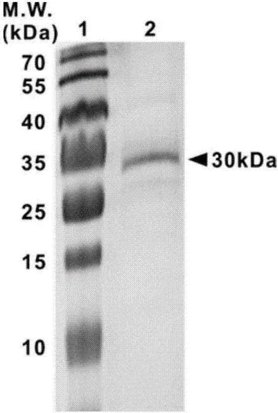
[0027] Example 1 Expression and purification of APEX2 protein

[0028] The pTRC99A-APEX2 plasmid (Addgene plasmid #72558) was purchased from addgene. The sequence of the pTRC99A-APEX2 plasmid is shown in SEQ ID No:1.

[0029] BL21-DE3 Escherichia coli competent cells were purchased from Tiangen Biochemical Technology (Beijing) Co., Ltd. Add 50ng of pTRC99A-APEX2 plasmid to competent cells, place on ice for 30min, heat shock at 42°C, add LB medium, culture at 37°C, 150rpm for 1h, take an appropriate amount of bacterial liquid and spread it on an ampicillin-resistant LB agarose plate, and culture overnight. A single clone was picked, inoculated into 1 mL ampicillin-resistant LB medium, and cultured overnight at 37°C and 220 rpm. Inoculate 1 mL of bacterial liquid into 500 mL of ampicillin-resistant LB medium, and incubate at 37°C, 220 rpm for about 5 hours. When the OD value of the bacterial liquid reaches 0.6, add 420 μM IPTG and 1 mM 5-aminolevulinic acid, at 18°C, 220 rpm ...

Embodiment 2

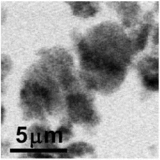
[0031] Example 2 Preparation of DAB Polymer and Synchronous X-ray Imaging Observation

[0032] DAB aqueous solution and H 2 o 2 respectively added to PBS buffer at pH 7.4, the final concentration of DAB was 0.4mg / mL, H 2 o 2 The final concentration is 10mM, mix well. APEX2 protein was added at a final concentration of 100 nM. After reacting for 15 minutes, the resulting DAB polymer suspension was dropped on the silicon nitride window.

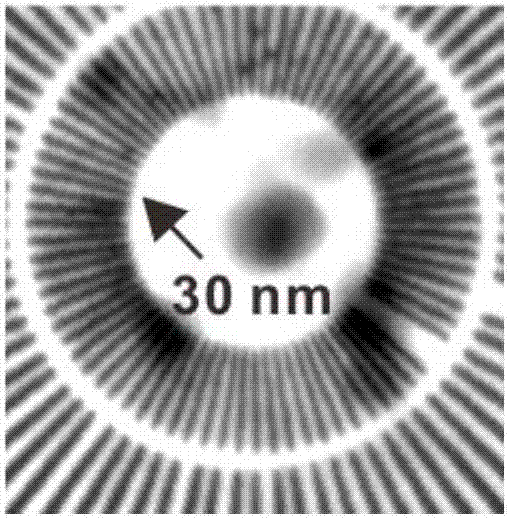
[0033]The X-ray imaging experiment of DAB polymer was carried out at the BL08U1 soft X-ray spectroscopy microscope station of Shanghai Light Source, and the experimental method was soft X-ray transmission imaging. The X-rays are extracted by the undulator, monochromatized by the planar grating monochromator, and then focused onto the sample by the zone plate, and then the transmitted photons are detected by the fast proportional counting detector PMT. The photon energy range is 250-2000eV, and the spatial resolution is 30nm. The DAB poly...

Embodiment 3

[0035] Example 3 APEX2, HRP, miniSOG and 700DX Catalyzed DAB Molecule to Generate Polymer and Its Application Comparison in Synchronous X-ray Imaging

[0036] APEX2 catalyzes DAB molecules to form polymers in vitro, and the method is the same as in Example 2.

[0037] HRP catalyzes DAB molecules to form polymers in vitro, and the method is as follows: HRP is purchased from Sigma (product number: P8375). DAB aqueous solution and H 2 o 2 respectively added to PBS buffer at pH 7.4, the final concentration of DAB was 0.4mg / mL, H 2 o 2 The final concentration is 10mM, mix well. HRP was added to a final concentration of 100 nM. The DAB polymer was collected after 15 min of reaction.

[0038] miniSOG catalyzes DAB molecules to generate polymers in vitro. The method is: clone the cDNA sequence of miniSOG into the pBAD-Myc-His A prokaryotic expression vector backbone to construct the pBAD-miniSOG plasmid. The sequence of the pBAD-miniSOG plasmid is shown in SEQ ID No:2. Add 5...


PUM


Abstract

The invention provides a preparation method of a synchronous X-ray visible imaging tag. The preparation method comprises the following steps: (1) dissolving substrate molecules in a buffer system; (2) adding enzyme or micromolecules having catalytic activity for the substrate molecules, and uniformly mixing; (3) reacting for a period of time until a polymer is generated; and (4) dropwise adding the suspension containing the polymer onto a synchronous imaging substrate, and synchronizing the X-ray imaging observation. The invention also provides a synchronously radiated X-ray visible imaging tag prepared according to the preparation method. According to the imaging tag, the X rays have the characteristics of good energy resolution, by virtue of in vitro chemical catalytic reaction, the synchronous X-ray visible imaging tag is successfully prepared, and good foundation is set for further preparing an X-ray sensitive molecular probe and realizing the specific identification and imaging of intracellular biomolecules.

Description

technical field [0001] The invention relates to the technical field of biochemistry, in particular to a synchronous X-ray visible imaging label and a preparation method thereof. Background technique [0002] Microscopic imaging technology is one of the main driving forces for the development of cell life science. Microscopy based on synchrotron X-rays has unique advantages in the field of cell imaging. Since the wavelength of X-rays is in the range of 0.1-10nm, it is naturally a super-resolution microscopic imaging technology, and the resolution can theoretically reach several nanometers. In addition, compared with electron beams, X-rays are more penetrating to biological samples, so intact cells can be imaged without processing such as slicing. More importantly, X-ray microscopic imaging technology has good energy resolution and can accurately distinguish the absorption spectra of many elements. Therefore, combined with X-ray-sensitive imaging probes, high-resolution rec...

Claims


Application Information

Patent Timeline
IPC IPC(8): G01N23/04
CPCG01N23/043
Inventor 樊春海诸颖孔华庭张继超夏凯闫庆龙王丽华胡钧
Owner SHANGHAI INST OF APPLIED PHYSICS - CHINESE ACAD OF SCI
Why Eureka
Try Eureka