Decellularized pulmonary support and preparation method thereof

A technology of decellularization and dodecyl ether, applied in the field of decellularized lung scaffolds and its preparation, can solve the problems of severe respiratory membrane rupture, restriction of lung transplantation, immune rejection, etc., and achieve good cellularization and vascularization, Conducive to quantitative production and unified preparation methods

Inactive Publication Date: 2017-08-29
FUWAI HOSPITAL CHINESE ACAD OF MEDICAL SCI & PEKING UNION MEDICAL COLLEGE
View PDF5 Cites 11 Cited by
  • Summary
  • Abstract
  • Description
  • Claims
  • Application Information

AI Technical Summary

Problems solved by technology

However, due to the serious shortage of donors, immune rejection, disease transmission, and low long-term survival rate, the development of lung transplantation is restricted.
At present, there are only less than 3,700 lung transplants in the world every year, which is far from meeting the demand
It is difficult for the commonly used detergents to fully remove the cells in the lungs while perfectly retaining the three-dimensional structure of the lungs
Excessive residual cells can lead to severe immun

Method used


Image

  • Decellularized pulmonary support and preparation method thereof
  • Decellularized pulmonary support and preparation method thereof
  • Decellularized pulmonary support and preparation method thereof

Examples


Embodiment 1

[0055] 1. Rat lung extraction

[0056] Select healthy male clean-grade SD rats, weighing 250-350g, aged 8-12 weeks, fasting for 12 hours and drinking water for 4 hours before the operation. Intraperitoneal injection of heparin, 1000U / Kg, 15min later intraperitoneal injection of 3% pentobarbital solution, the dosage is 150mg / Kg for anesthesia. After successful anesthesia, the rats were placed in a supine position, and the limbs were fixed on the operating table with thumbtacks. Thoracic and abdominal surgical areas were prepared for skin preparation and routinely disinfected. A combined midline incision was made in the chest and abdomen, about 5-8 cm in length. The diaphragm and ribs were cut open, and the thymus was removed to expose the heart and lungs. The abdominal viscera were removed with a cotton swab, and the abdominal aorta and main vein were cut off to let blood out. Inject 20-50ml of normal saline through the right ventricle. Each 1ml of normal saline contains 50U ...

experiment example 1

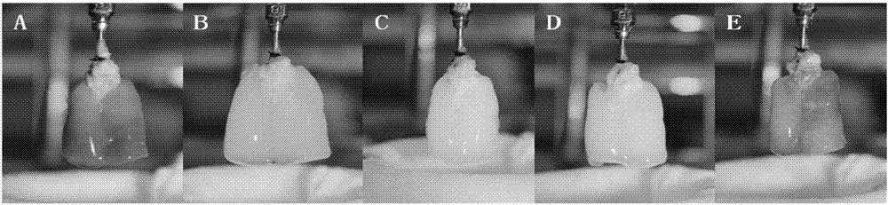
[0068] Different perfusion methods were used to observe the lung status of rats, such as figure 1 As shown, A is a normal rat lung taken out of the refrigerator, separated from the heart, inserted into a 16G perfusion head through the main pulmonary artery, and suspended to the Langendorff operating table to wait for perfusion. B is perfusion with 0.1% SLES solution at a flow rate of 5ml / min. After 2 hours, the lungs are basically transparent. In order to achieve the effect of basic transparency, the perfusion time can be extended to 3 hours. C is 1 hour after infusion of DNase I solution with a concentration of 30ug / ml, the flow rate is 2ml / min, which makes the lungs more crystal clear. D is the decellularized lung scaffold prepared by the present invention after the perfusion is finally completed. After perfusing the methylene blue dye solution into the stent prepared by the method of the E researcher, it can be seen that the blue dye solution is evenly distributed and the ...

experiment example 2

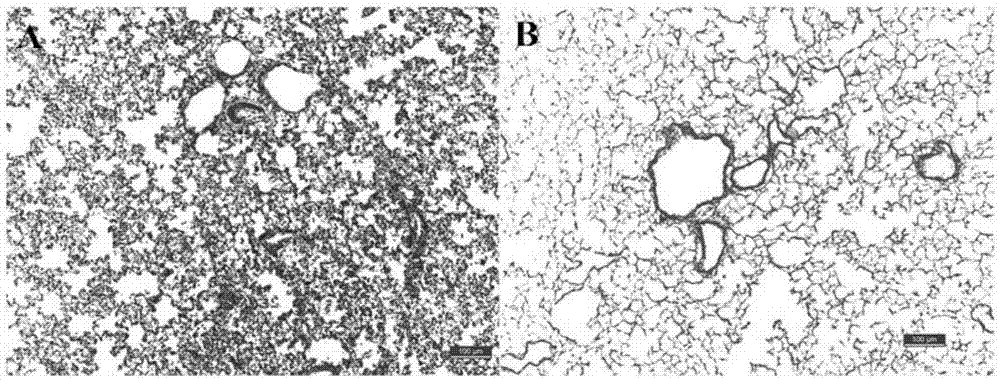
[0070] figure 2 It is the comparison chart of HE staining between normal rat lung tissue and the lung scaffold prepared by the present invention; normal lung lobes and decellularized lung scaffold lung lobes were randomly selected according to the conventional HE staining steps, that is, routine HE staining after fixation, dehydration, embedding, and preparation of slices. Such as figure 2 As shown, A is the HE staining of normal rat lung tissue, and the tissue is full of cells; B is the HE staining of the lung scaffold prepared by the present invention, the tissue has no obvious cell distribution, magnified 100 times, and the scale bar is 100um. pass figure 2 It can be concluded that the method of the present invention can completely remove normal lung cells.


PUM


Abstract

The invention provides a decellularized pulmonary support. The decellularized pulmonary support is prepared from sodium lauryl ether sulfate and a DNase I solution through injection treatment. The decellularized pulmonary support prepared by a langendorff injection system is clean in cell removal, a three-dimensional structure and an extracellular matrix protein are reserved completely, and the fusion, cellularization and vascularization are good 6 weeks after allograft subcutaneous transplanting. A preparation method of the decellularized pulmonary support is high in efficiency and simple in step, can be applied to batch production and has a good application prospect in the fields of tissue engineering lungs and loose organs.

Description

technical field [0001] The invention relates to the field of tissue engineering artificial lungs, in particular to a decellularized lung scaffold and a preparation method thereof. Background technique [0002] At present, due to environmental factors, genetic factors, personal factors and other reasons, chronic obstructive pulmonary disease (COPD), idiopathic pulmonary fibrosis (IPF), ɑ-1 antitrypsin deficiency, pulmonary hypertension and other lung diseases are more and more common. , for these patients, lung transplantation is the only sure-fire treatment. However, due to severe shortage of donors, immune rejection, disease transmission, low long-term survival rate and other reasons, the development of lung transplantation is restricted. At present, there are only less than 3,700 lung transplants worldwide each year, which is far from meeting the demand. With the development of tissue engineering and regenerative medicine, especially the successful clinical application o...

Claims


Application Information

Patent Timeline
IPC IPC(8): A61L27/36
CPCA61L27/3604A61L27/3641A61L27/3687A61L2430/40
Inventor 黑飞龙马金辉鞠志海于洁乔叶薷候陈玮王晨陈书弘罗富良
Owner FUWAI HOSPITAL CHINESE ACAD OF MEDICAL SCI & PEKING UNION MEDICAL COLLEGE
Why Eureka
Try Eureka